

Characterization of cardiac time intervals in healthy bonnet macaques (Macaca radiata) by using an electronic stethoscope
- PMID: 21439218
- PMCID: PMC3061425
Characterization of cardiac time intervals in healthy bonnet macaques (Macaca radiata) by using an electronic stethoscope
Abstract
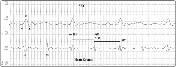
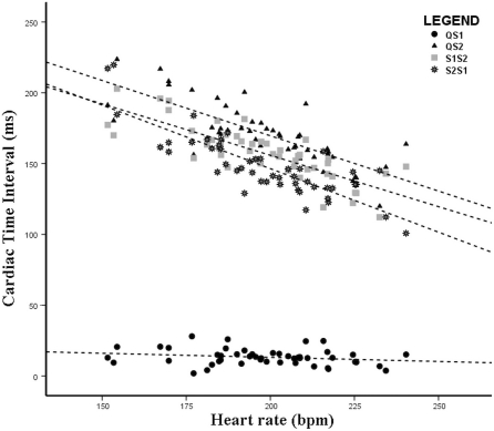
Nonhuman primates are used frequently in cardiovascular research. Cardiac time intervals derived by phonocardiography have long been used to assess left ventricular function. Electronic stethoscopes are simple low-cost systems that display heart sound signals. We assessed the use of an electronic stethoscope to measure cardiac time intervals in 48 healthy bonnet macaques (age, 8±5 y) based on recorded heart sounds. Technically adequate recordings were obtained from all animals and required 1.5±1.3 min. The following cardiac time intervals were determined by simultaneously recording acoustic and single-lead electrocardiographic data: electromechanical activation time (QS1), electromechanical systole (QS2), the time interval between the first and second heart sounds (S1S2), and the time interval between the second and first sounds (S2S1). QS2 was correlated with heart rate, mean arterial pressure, diastolic blood pressure, and left ventricular ejection time determined by using echocardiography. S1S2 correlated with heart rate, mean arterial pressure, diastolic blood pressure, left ventricular ejection time, and age. S2S1 correlated with heart rate, mean arterial pressure, diastolic blood pressure, systolic blood pressure, and left ventricular ejection time. QS1 did not correlate with any anthropometric or echocardiographic parameter. The relation S1S2/S2S1 correlated with systolic blood pressure. On multivariate analyses, heart rate was the only independent predictor of QS2, S1S2, and S2S1. In conclusion, determination of cardiac time intervals is feasible and reproducible by using an electrical stethoscope in nonhuman primates. Heart rate is a major determinant of QS2, S1S2, and S2S1 but not QS1; regression equations for reference values for cardiac time intervals in bonnet macaques are provided.
Figures
References
-
- Abelson D, Kamens EA. 1977. Bedside measurement of systolic and diastolic time intervals using the stethometer. Cardiovasc Res 11:270–274 - PubMed
-
- Abrahams C, Janicki JS, Weber KT. 1987. Myocardial hypertrophy in Macaca fascicularis. Structural remodeling of the collagen matrix. Lab Invest 56:676–683 - PubMed
-
- Bombardini T, Gemignani V, Bianchini E, Venner L, Petersen C, Pasanisi E, Pratali L, Alonso-Rodriguez D, Mascia Pianelli M, Francesco Faita F, Giannoni M, Arpesella G, Picano E. 2008. Diastolic time–frequency relation in the stress echo lab: filling timing and flow at different heart rates. Cardiovasc Ultrasound 6:15. - PMC - PubMed
-
- Buss DD, Hyde DM, Poulos PW., Jr 1982. Coronary artery distribution in bonnet monkeys (Macaca radiata). Anat Rec 203:411–417 - PubMed
-
- Collins SP, Lindsell CJ, Kontos MC, Zuber M, Kipfer P, Attenhofer Jost C, Kosmicki D, Michaels AD. 2009. Bedside prediction of increased filling pressure using acoustic electrocardiography. Am J Emerg Med 27:397–408 - PubMed
Publication types
MeSH terms
Substances
LinkOut - more resources
Full Text Sources