

Primary hepatic Mycobacterium tuberculosis complex infection with terminal dissemination in a pig-tailed macaque (Macaca nemestrina)
- PMID: 21439222
- PMCID: PMC3061429
Primary hepatic Mycobacterium tuberculosis complex infection with terminal dissemination in a pig-tailed macaque (Macaca nemestrina)
Abstract
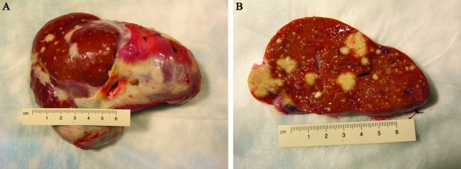
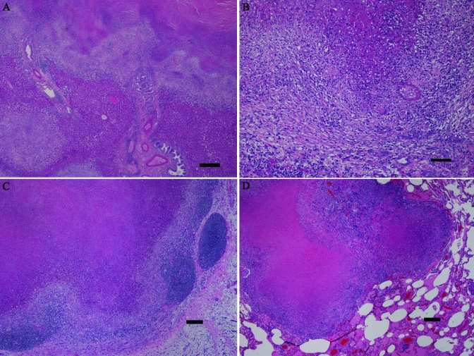
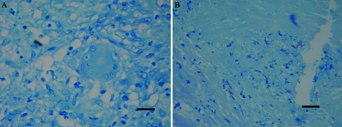
An adult, female, pig-tailed macaque (Macaca nemestrina) of Indonesian origin presented with profound weight loss, anemia (PCV, 29%; normal, 36% to 45%), hypoalbuminemia (1.0 g/dL; normal, 3.5 to 5.2 g/dL), elevated alkaline phosphatase (1990 U/L; normal, 26 to 98 U/L), and an elevated erythrocyte sedimentation rate (75 mm/h; normal, less than 20 mm/h). Abdominal ultrasonography demonstrated an enlarged liver with hyperechoic areas. Euthanasia was performed. Grossly, the liver had multifocal, effacing, white masses throughout and was enlarged with rounded edges. There were 2, small nodules in the right lung lobes. Histologically, the hepatic masses were densely fibrous-encapsulated granulomas with vast central necrosis. The lung nodules also were maturing granulomas, and one kidney and one atrium had small, early granulomas. Fite acid-fast stains of liver and lung revealed very few acid-fast bacilli. PCR analysis of paraffin-embedded liver identified Mycobacterium tuberculosis complex. Culture of the liver was negative twice. This macaque had 16 negative intradermal tuberculin skin tests over the course of 6 y. We hypothesize that the animal arrived with a latent hepatic or enteric infection that later recrudesced and disseminated. Primary hepatic mycobacteriosis is not a typical presentation of tuberculosis in macaques. Negative tuberculin skin tests can be seen with latent infections and extrapulmonary tuberculosis such as Pott disease. This case underscores the problems associated with current surveillance procedures and the risks associated with latent mycobacterial infections in macaques.
Figures
References
-
- Alvarez S, McGabe WR. 1984. Extrapulmonary tuberculosis revisited: a review of experience at Boston City and other hospitals. Medicine (Baltimore) 63:25–55 - PubMed
-
- Animal Welfare Act as Amended 2007. 7 USC §2131–2156.
-
- Bernacky BJ, Gibson SV, Keeling ME, Abee CR. 2002. Nonhuman primates, p 676–777 In: Fox JG, Anderson LC, Loew FM. Laboratory animal medicine, 2nd ed San Diego (CA): Academic Press
-
- Clingerman KJ, Summers L. 2005. Development of a body condition scoring system for nonhuman primates using Macaca mulatta as a model. Lab Anim (NY) 34:31–36 - PubMed
-
- de Sanctis JT, Carpenter C, Sims M, SenGupta D, Prentice JL, Cookson BT, Boyanton BL. 2010. Culture-negative endocarditis and the use of molecular diagnostics. A case report. Infect Dis Clin Pract 18:120–123
Publication types
MeSH terms
Grants and funding
LinkOut - more resources
Full Text Sources
Medical
Research Materials
Miscellaneous