Review
. 2011 Jun;94(1):1-19.
doi: 10.1016/j.pneurobio.2011.03.005. Epub 2011 Mar 23.

Gene-environment interactions: key to unraveling the mystery of Parkinson's disease

Affiliations
Review

Gene-environment interactions: key to unraveling the mystery of Parkinson's disease

Hui-Ming Gao et al. Prog Neurobiol. 2011 Jun.

Abstract

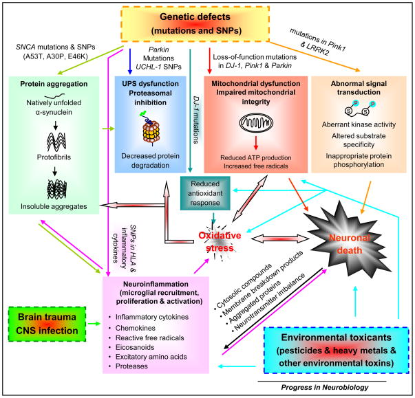
Parkinson's disease (PD) is the second most common neurodegenerative disease. The gradual, irreversible loss of dopamine neurons in the substantia nigra is the signature lesion of PD. Clinical symptoms of PD become apparent when 50-60% of nigral dopamine neurons are lost. PD progresses insidiously for 5-7 years (preclinical period) and then continues to worsen even under the symptomatic treatment. To determine what triggers the disease onset and what drives the chronic, self-propelling neurodegenerative process becomes critical and urgent, since lack of such knowledge impedes the discovery of effective treatments to retard PD progression. At present, available therapeutics only temporarily relieve PD symptoms. While the identification of causative gene defects in familial PD uncovers important genetic influences in this disease, the majority of PD cases are sporadic and idiopathic. The current consensus suggests that PD develops from multiple risk factors including aging, genetic predisposition, and environmental exposure. Here, we briefly review research on the genetic and environmental causes of PD. We also summarize very recent genome-wide association studies on risk gene polymorphisms in the emergence of PD. We highlight the new converging evidence on gene-environment interplay in the development of PD with an emphasis on newly developed multiple-hit PD models involving both genetic lesions and environmental triggers.


Figures

Figure 1
Figure 1. PD develops from complex gene-environment interactions and involves multiple molecular pathways that crosstalk and converge leading to PD neurodegeneration
The identification of causative gene defects in PD has defined four major pathways leading to neuronal demise: protein aggregation, ubiquitin-proteasome system (UPS) dysfunction and impaired protein degradation, mitochondrial dysfunction and integrity impairment, and aberrant signal transduction. SNCA mutations and single-nucleotide polymorphisms (SNPs) make α-synuclein adopt a propensity for misfolding and accelerated aggregate formation. Excessive α-synuclein aggregates may overwhelm UPS protein degeneration. Accumulated α-synuclein can translocate to the mitochondria and impair mitochondrial activity. Parkin mutations and UCHL-1 SNPs prevent the proteolytic degradation of excessive toxic proteins (e.g. misfolded α-synuclein) in proteasomal machinery. PINK1, Parkin, and DJ-1 functionally interact to maintain mitochondrial integrity and functionality and to protect cells against adverse effects of multiple stressors. Mutations in these genes cause mitochondrial dysfunction and subsequent decline in ATP production and increase in free radical generation, which results in oxidative stress and energy deficiency. Impaired mitochondria can release cytochrome c and other ‘pro-apoptotic factors’ triggering apoptotic cascades and cell death. Mitochondria in at least some forms of PD reveal abnormal morphology, impaired fission-fusion balance, and metabolic malfunction. DJ-1 mutations reduce antioxidant response of cells, aggravating oxidative stress. Oxidative stress engages in diverse cellular processes and plays a prominent role in the induction of neuronal death. For instance, excessive production of free radicals can damage proteins (e.g. abnormal modification of α-synuclein and inactivation of Parkin), lipids, DNA, or RNA, leading to cell dysfunction (e.g. UPS and mitochondrial impairment) and eventual death. Mutations in Pink1 and LRRK2 induced aberrant kinase activity, altered substrate specificity, leading to inappropriate protein phosphorylation (e.g. increased α-synuclein phosphorylation at serine 129 by LRRK2 in vitro) and thereby affecting cell survival. Environmental toxins and brain trauma can trigger neuronal lesions by damaging mitochondria, causing oxidative stress, inducing inflammation in the central nervous system (CNS), and compromising defence mechanisms of cells. Some environmental risk factors can directly activate microglia (the resident immune cells in the CNS) or cause systemic inflammation, which in turn affects CNS inflammation. Genetic variation and polymorphisms in the HLA region and several inflammatory cytokines may become risk factors for PD. Activated microglia produce and secrete a spectrum of inflammatory and cytotoxic molecules, such as cytokines, chemokines, reactive free radicals, eicosanoids, and proteases. In addition to modulating microglial activity, these molecules influence the fate of surrounding neurons. Excessive inflammatory reaction usually becomes exaggerated and destructive, and turns into chronic inflammation that drives progressive neurodegenerative process. Injured neurons activate the surrounding microglia through the release or leakage of noxious self-compounds into the extracellular milieu, such as membrane breakdown products, abnormally processed or aggregated proteins (e.g. α-synuclein and β-amyloid), imbalanced neurotransmitters (e.g. elevated glutamate) and cytosolic compounds (e.g. α-synuclein, ATP, HMGB1 and neuromelanin). Thus, gene-environment interplay induces complex crosstalk among multiple signal cascades, forming a network and culminating in neuronal death and PD development.

References

    1. Abeliovich A, Schmitz Y, Farinas I, Choi-Lundberg D, Ho WH, Castillo PE, Shinsky N, Verdugo JM, Armanini M, Ryan A, Hynes M, Phillips H, Sulzer D, Rosenthal A. Mice lacking alpha-synuclein display functional deficits in the nigrostriatal dopamine system. Neuron. 2000;25:239–252. - PubMed
    1. Ahn TB, Kim SY, Kim JY, Park SS, Lee DS, Min HJ, Kim YK, Kim SE, Kim JM, Kim HJ, Cho J, Jeon BS. alpha-Synuclein gene duplication is present in sporadic Parkinson disease. Neurology. 2008;70:43–49. - PubMed
    1. Alam M, Schmidt WJ. Rotenone destroys dopaminergic neurons and induces parkinsonian symptoms in rats. Behav Brain Res. 2002;136:317–324. - PubMed
    1. Andres-Mateos E, Mejias R, Sasaki M, Li X, Lin BM, Biskup S, Zhang L, Banerjee R, Thomas B, Yang L, Liu G, Beal MF, Huso DL, Dawson TM, Dawson VL. Unexpected lack of hypersensitivity in LRRK2 knock-out mice to MPTP (1-methyl-4-phenyl-1,2,3,6-tetrahydropyridine) J Neurosci. 2009;29:15846–15850. - PMC - PubMed
    1. Andres-Mateos E, Perier C, Zhang L, Blanchard-Fillion B, Greco TM, Thomas B, Ko HS, Sasaki M, Ischiropoulos H, Przedborski S, Dawson TM, Dawson VL. DJ-1 gene deletion reveals that DJ-1 is an atypical peroxiredoxin-like peroxidase. Proc Natl Acad Sci U S A. 2007;104:14807–14812. - PMC - PubMed

Publication types