. 2011 Mar 25;18(3):284-91.
doi: 10.1016/j.chembiol.2011.01.014.

Lipidomics at the interface of structure and function in systems biology

Affiliations

Lipidomics at the interface of structure and function in systems biology

Richard W Gross et al. Chem Biol. .

Abstract

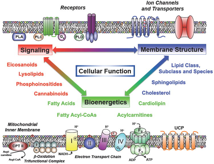
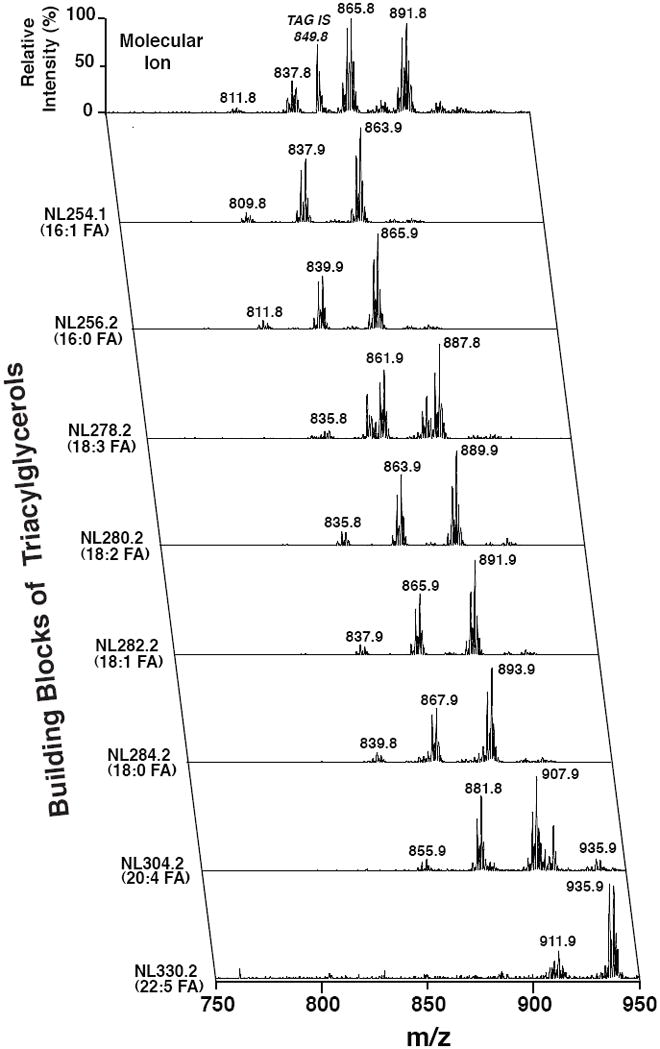
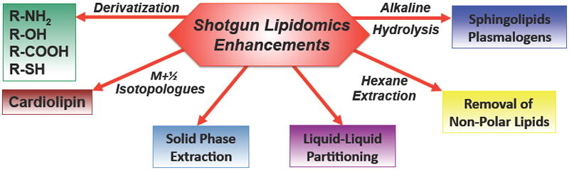
Cells, tissues, and biological fluids contain a diverse repertoire of many tens of thousands of structurally distinct lipids that play multiple roles in cellular signaling, bioenergetics, and membrane structure and function. In an era where lipid-related disease states predominate, lipidomics has assumed a prominent role in systems biology through its unique ability to directly identify functional alterations in multiple lipid metabolic and signaling networks. The development of shotgun lipidomics has led to the facile accrual of high density information on alterations in the lipidome mediating physiologic cellular adaptation during health and pathologic alterations during disease. Through both targeted and nontargeted investigations, lipidomics has already revealed the chemical mechanisms underlying many lipid-related disease states.


Figures

Figure 1
Figure 1. The Pleiotropic Roles of Lipids in Cellular Function
Lipids fulfill multiple roles in cellular function including cellular signaling (top left) through:1) harboring latent 2nd messengers of signal transduction that are released by phospholipases (PLA, PLC and PLD enzymes); 2) covalent transformation of membrane lipids into biologically active ligands by kinases (e.g., PI 3,4,5 triphosphate); 3) providing molecular scaffolds for the assembly of protein complexes mediating receptor/effector coupling (e.g., G-protein coupled receptors); and 4) coupling the vibrational, rotational and translational energies and dynamics of membrane lipids to transmembrane proteins such as ion channels and transporters (top right) thereby facilitating dynamic cooperative lipid–protein interactions that collectively regulate transmembrane protein function. Moreover, lipids play essential roles in mitochondrial cellular bioenergetics (bottom) through the use of fatty acids as substrates for mitochondrial β-oxidation (bottom left) that result in the production of reducing equivalents (e.g., NADH). The chemical energy in NADH is harvested through oxidative phosphorylation whose flux is tightly regulated by mitochondrial membrane constituents including cardiolipins which modulate electron transport chain (ETC) supercomplex formation. A second mechanism modulating mitochondrial energy production is the dissipation of the proton gradient by the transmembrane flip-flop of fatty acids in the mitochondrial inner membrane bilayer as well as the fatty acid-mediated regulation of uncoupling proteins (UCP).
Figure 2
Figure 2. Electrospray Ionization Mass Spectrometric Analysis of Extracts of Murine White Adipose Tissue by MDMS-SL
Bligh and Dyer extracts of white adipose tissue from mice fed a high fat diet were prepared as previously described (Han and Gross, 2005) and directly infused into the ESI ion source. Positive-ion ESI mass spectra identified multiple molecular ions in the full mass scan (top row; x-axis) that were quantitated through ratiometric comparisons with internal standard (tri 17:1 triacylglycerol, m/z 849.8) after corrections for isotope abundance and acyl chain length and unsaturation effects. Tandem mass spectrometric analysis was performed using neutral loss scanning for the indicated naturally occurring aliphatic chains. All scans were normalized to the base peak of the individual spectrum. Through bioinformatic analysis of the ion counts in each row (x-axis scan) and column (y-axis scans), the compositional identities of each individual molecular species can be determined and their relative abundance can be quantified.
Figure 3
Figure 3. Enhanced Shotgun Lipidomics Approaches
The analytic power of shotgun lipidomics can be extended through exploiting the unique chemical characteristics of specific lipid classes. These include the derivatization of nucleophilic lipid moieties to increase signal intensity and/or engender a mass shift to facilitate mass spectrometric analyses, the use of a M+1/2 isotopologue approach for doubly negatively charged cardiolipins, multiplexed extractions including liquid/liquid partitioning, liquid/solid partitioning (solid phase extraction) and/or alkaline hydrolysis to enrich for sphingolipids or ether lipids. In some tissues (e.g., adipose tissue), the removal of non-polar lipids by hexane extraction is judicious prior to the subsequent analysis of polar lipid constituents.

References

    1. Bartz R, Li W-H, Venables B, Zehmer JK, Roth MR, Welti R, Anderson RG, Liu P, Chapman KD. Lipidomics reveals that adiposomes store ether lipids and mediate phospholipid traffic. J Lipid Res. 2007;48:837–847. - PubMed
    1. Beckonert O, Coe M, Keun HC, Wang Y, Ebbels TMD, Holmes E, Lindon JC, Nicholson JK. High-resolution magic-angle-spinning NMR spectroscopy for metabolic profiling of intact tissues. Nat Protoc. 2010;5:1019–1032. - PubMed
    1. Bollinger JG, Thompson W, Lai Y, Oslund RC, Hallstrand TS, Sadilek M, Turecek F, Gelb MH. Improved sensitivity mass spectrometric detection of eicosanoids by charge reversal derivatization. Anal Chem. 2010;82:6790–6796. - PMC - PubMed
    1. Breen EP, Gouin SG, Murphy AF, Haines LR, Jackson AM, Pearson TW, Murphy PV, Porter RK. On the mechanism of mitochondrial uncoupling protein 1 function. J Biol Chem. 2005;281:2114–2119. - PubMed
    1. Cady SD, Schmidt-Rohr K, Wang J, Soto CS, DeGrado WF, Hong M. Structure of the amantadine binding site of influenza M2 proton channels in lipid bilayers. Nature. 2010;463:689–693. - PMC - PubMed

Publication types

LinkOut - more resources