

Multiscale imaging characterization of dopamine transporter knockout mice reveals regional alterations in spine density of medium spiny neurons
- PMID: 21439946
- PMCID: PMC3104025
- DOI: 10.1016/j.brainres.2011.03.044
Multiscale imaging characterization of dopamine transporter knockout mice reveals regional alterations in spine density of medium spiny neurons
Abstract
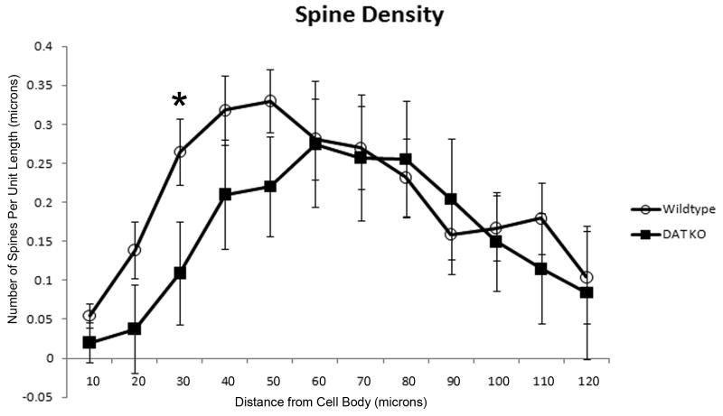
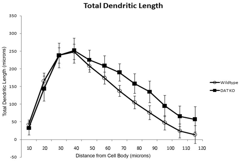
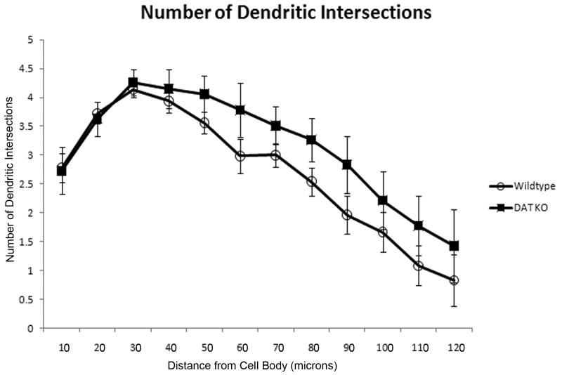
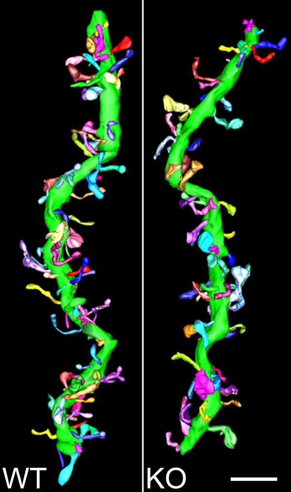
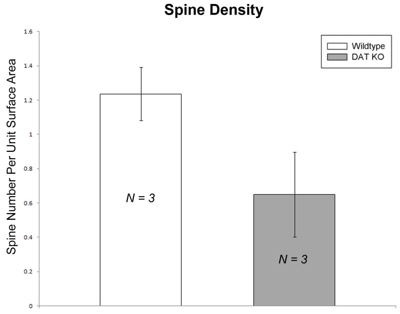
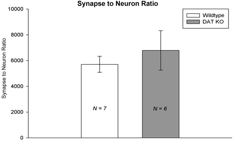
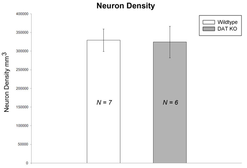
The dopamine transporter knockout (DAT KO) mouse is a model of chronic hyperdopaminergia used to study a wide range of neuropsychiatric disorders such as schizophrenia, attention deficit hyperactivity disorder (ADHD), drug abuse, depression, and Parkinson's disease (PD). Early studies characterizing this mouse model revealed a subtle, but significant, decrease in the anterior striatal volume of DAT KO mice accompanied by a decrease in neuronal cell body numbers (Cyr et al., 2005). The present studies were conducted to examine medium spiny neuron (MSN) morphology by extending these earlier reports to include multiscale imaging studies using correlated light microscopy (LM) and electron microscopy (EM) techniques. Specifically, we set out to determine if chronic hyperdopaminergia results in quantifiable or qualitative changes in DAT KO mouse MSNs relative to wild-type (WT) littermates. Using Neurolucida Explorer's morphometric analysis, we measured spine density, dendritic length and synapse number at ages that correspond with the previously reported changes in striatal volume and progressive cell loss. Light microscopic analysis using Neurolucida tracings of photoconverted striatal MSNs revealed a highly localized loss of dendritic spines on the proximal portion of the dendrite (30 μm from the soma) in the DAT KO group. Next, thick sections containing MSN dendritic segments located at a distance of 20-60 μm from the cell soma, a region of the dendrite where spine density is reported to be the highest, were analyzed using electron microscope tomography (EMT). Because of the resolution limits of LM, the EM analysis was an extra measure taken to assure that our analysis included nearly all spines. Spine density measurements collected from the EMT data revealed only a modest decrease in the DAT KO group (n=3 mice) compared to age-matched WT controls (n=3 mice), a trend that supports the LM findings. Finally, a synaptic quantification using unbiased stereology did not detect a difference between DAT KO mice (n=6 mice) and WT controls (n=7 mice) at the EM level, supporting the focal nature of the early synaptic loss. These findings suggest that DAT KO mice have MSNs with highly localized spine loss and not an overall morphologically distinct cell shape. The characterization of morphological changes in DAT KO mice may provide information about the neural substrates underlying altered behaviors in these mice, with relevance for human neurological disorders thought to involve altered dopaminergic homeostasis. Results from this study also indicate the difficulty in correlating structural changes across scales, as the results on fine structure revealed thus far are subtle and non-uniform across striatal MSNs. The complexities associated with multiscale studies are driving the development of shared online informatics resources by gaining access to data where it is being analyzed.
Copyright © 2011 Elsevier B.V. All rights reserved.
Figures

References
-
- Allen PB, Zachariou V, Svenningsson P, Lepore AC, Centonze D, Costa C, Rossi S, Bender G, Chen G, Feng J, Snyder GL, Bernardi G, Nestler EJ, Yan Z, Calabresi P, Greengard P. Distinct roles for spinophilin and neurabin in dopamine-mediated plasticity. Neuroscience. 2006;140:897–911. - PubMed
-
- Cyr M, Caron MG, Johnson GA, Laakso A. Magnetic resonance imaging at microscopic resolution reveals subtle morphological changes in a mouse model of dopaminergic hyperfunction. Neuroimage. 2005;26:83–90. - PubMed
Publication types
MeSH terms
Substances
Grants and funding
LinkOut - more resources
Full Text Sources
Molecular Biology Databases
Research Materials
Miscellaneous
