. 2011 Jul;141(1):358-69.
doi: 10.1053/j.gastro.2011.03.041. Epub 2011 Mar 24.

TLR9 and the NLRP3 inflammasome link acinar cell death with inflammation in acute pancreatitis

Affiliations

TLR9 and the NLRP3 inflammasome link acinar cell death with inflammation in acute pancreatitis

Rafaz Hoque et al. Gastroenterology. 2011 Jul.

Abstract

Background & aims: Acute pancreatitis is characterized by early activation of intracellular proteases followed by acinar cell death and inflammation. Activation of damage-associated molecular pattern (DAMP) receptors and a cytosolic complex termed the inflammasome initiate forms of inflammation. In this study, we examined whether DAMP-receptors and the inflammasome provide the link between cell death and the initiation of inflammation in pancreatitis.

Methods: Acute pancreatitis was induced by caerulein stimulation in wild-type mice and mice deficient in components of the inflammasome (apoptosis-associated speck-like protein containing a caspase recruitment domain [ASC], NLRP3, caspase-1), Toll-like receptor 9 (TLR9), or the purinergic receptor P2X(7). Resident and infiltrating immune cell populations and pro-interleukin-1β expression were characterized in control and caerulein-treated adult murine pancreas. TLR9 expression was quantified in pancreatic cell populations. Additionally, wild-type mice were pretreated with a TLR9 antagonist before induction of acute pancreatitis by caerulein or retrograde bile duct infusion of taurolithocholic acid 3-sulfate.

Results: Caspase-1, ASC, and NLRP3 were required for inflammation in acute pancreatitis. Genetic deletion of Tlr9 reduced pancreatic edema, inflammation, and pro-IL-1β expression in pancreatitis. TLR9 was expressed in resident immune cells of the pancreas, which are predominantly macrophages. Pretreatment with the TLR9 antagonist IRS954 reduced pancreatic edema, inflammatory infiltrate, and apoptosis. Pretreatment with IRS954 reduced pancreatic necrosis and lung inflammation in taurolithocholic acid 3-sulfate-induced acute pancreatitis.

Conclusions: Components of the inflammasome, ASC, caspase-1, and NLRP3, are required for the development of inflammation in acute pancreatitis. TLR9 and P2X(7) are important DAMP receptors upstream of inflammasome activation, and their antagonism could provide a new therapeutic strategy for treating acute pancreatitis.


Figures

Figure 1
Figure 1
Deletion of caspase-1 (Casp1−/−) reduces the severity of acute pancreatitis. Acute pancreatitis in wild-type and Casp1−/− mice was induced by caerulein. Representative histology (A) and histology scores C) are shown. Gr1 immunohistochemistry (B) and GR1 positive cell counts in pancreatic tissue section (D) are shown. Asterisks denote significant differences (P<0.05) in bracketed groups. NS, not significant.
Figure 2
Figure 2
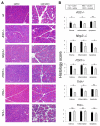
Deletion of inflammasome component ASC (ASC−/−), Nlrp3 (Nlrp3−/−), P2X7 receptor (P2X7−/−), or TOLL-like receptor 9 (Tlr9−/−) reduces the severity of acute pancreatitis. Acute pancreatitis in wild-type, ASC−/−, P2X7−/−, Tlr9−/−, and Tlr3−/− mice was induced by caerulein. Representative histology (A) is shown. Histology scores for pancreatic edema, inflammatory infiltrate, and apoptosis (B) are shown. Asterisks denote significant differences (P<0.05) in bracketed groups.
Figure 3
Figure 3
Genetic deletion of Tlr9 (Tlr9−/−) reduces pro-inflammatory cytokine production in caerulein-induced acute pancreatitis. Tlr9 mRNA is strongly and predominantly expressed in bone marrow-derived cells. Acute pancreatitis in wild-type and Tlr9−/− mice was induced by caerulein. One hour after the last injection of caerulein, pancreata were harvested and pancreatic pro-IL-1β mRNA was quantified by real-time PCR (A). Pancreatic cuboidal ductal cells (CD34 CD133+ CD45) and endothelial cells (CD34+ CD133 CD45) were isolated by flow cytometry (B). CD45+ bone marrow derived cells within the pancreas were assessed by flow cytometry for cell surface expression of tissue macrophage markers F4/80 and ER-MP23 (C and D). Tlr9 mRNA was quantified by real-time PCR from wild-type pancreatic subpopulations and expressed relative to whole spleen (E). Open bars: wild-type mice treated with saline. Solid bars: wild-type mice treated with caerulein. Gray bars: report results from Tlr9−/− mice treated with saline. Diagonal striped bars: Tlr9−/− mice treated with caerulein. Asterisks denote significant differences (P<0.05) in bracketed groups. # denotes significant difference (P<0.05) compared to all other bars shown.
Figure 4
Figure 4
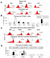
Macrophages are the predominant immune cell in the pancreas. The early inflammatory infiltrate in caerulein-induced acute pancreatitis is neutrophil predominant and genetic deletion of Tlr9 reduces this neutrophil infiltration. Pancreas and spleen were isolated from adult wild-type mice, single cell suspensions prepared, and flow cytometric analysis for cell surface proteins performed. Representative histograms for Gr1, F4/80, NK1.1, and CD3 cell surface expression in CD45+ bone marrow derived cells in the pancreas and spleen are shown (A). Resident immune cell populations are reported as percentage of total cells analyzed and as percentage of total CD45+ cells analyzed (B). Acute pancreatitis in wild-type and Tlr9−/− mice was induced by caerulein. Representative histograms (C) are shown for Gr1, F4/80, NK1.1, and CD3 cell surface expression in CD45+ pancreatic cells from wild-type and Tlr9−/− mice treated with caerulein. Resident immune cell populations are reported as percentage of total CD45+ cells analyzed (D). # denotes the predominant resident immune cell population within the pancreas. Asterisks denotes significant differences (P<0.05) between marked groups.
Figure 5
Figure 5
Pretreatment with the TLR9 antagonist IRS954 reduces the severity of caerulein-induced acute pancreatitis. Representative histology (A) and histology scores for pancreatic edema, inflammatory infiltrate, and apoptosis (D) are shown. Gr1 immunohistochemistry (B) and GR1 positive cell counts in pancreatic tissue section (C) are shown. TUNEL positivity (E) was scored in the groups above as well as in Casp1−/− mice subjected to caerulein pancreatitis. Pancreatic pro-IL-1β transcription is shown (F).
Figure 6
Figure 6
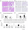
Pretreatment with the TLR9 antagonist reduces the severity of acute pancreatitis. Representative pancreatic head histology after hematoxylin and eosin staining (A) and histology scores for inflammatory infiltrate and necrosis (C) are shown. Representative lung histology after hematoxylin and eosin staining (B) and histology scores for lung edema and inflammatory infiltrate (D) are shown. Serum amylase is shown for both the TLCS and caerulein induced pancreatitis models (E). Asterisks denote significant differences (P<0.05) in bracketed groups. NS, not significant.
Figure 7
Figure 7
Pancreatic injury releases DAMPs. Circulating genomic DNA and mitochondrial DNA was quantitated in acute pancreatic injury by QPCR of serum for β-actin and ATP6 DNA (A). IL-1β release into supernatant was quantitated in peritoneal macrophages stimulated with pancreatic homogenate in the presence or absence of P2X7 antagonist A-438079 (B). NF-κB activation was quantitated by cell counts for immunochemically detected nuclear translocation in macrophages treated with pancreatic DNA or homogenate in the presence of TLR9 antagonist IRS954 or P2X7 antagonist A-438079, respectively (C). Representative imaging of nuclear localized NF-kB (D). Schematic representation of TLR9 and P2X7 mediated innate immune responses in acute pancreatic injury (E). Pancreatic acinar cell injury and necrosis results in release of DAMPs, including nuclear DNA (nDNA) and mitochondrial DNA (mtDNA) as well as intracellular ATP. Resident macrophages within the pancreas sense these DAMPs through (i) TLR9 receptors with respective induction of NF-κB translocation and pro-IL1 β transcription and through (ii) P2X7 with proteolytic maturation of IL-1β through the Nlrp3 inflammasome. IL-1β dependent pathways induce further pancreatic injury.

References

    1. O’Reilly DA, Kingsnorth AN. A brief history of pancreatitis. J R Soc Med. 2001;94:130–2. - PMC - PubMed
    1. Lampel M, Kern HF. Acute interstitial pancreatitis in the rat induced by excessive doses of a pancreatic secretagogue. Virchows Arch A Pathol Anat Histol. 1977;373:97–117. - PubMed
    1. Gravante G, Garcea G, Ong SL, Metcalfe MS, Berry DP, Lloyd DM, Dennison AR. Prediction of mortality in acute pancreatitis: a systematic review of the published evidence. Pancreatology. 2009;9:601–14. - PubMed
    1. Gukovskaya AS, Vaquero E, Zaninovic V, Gorelick FS, Lusis AJ, Brennan ML, Holland S, Pandol SJ. Neutrophils and NADPH oxidase mediate intrapancreatic trypsin activation in murine experimental acute pancreatitis. Gastroenterology. 2002;122:974–84. - PubMed
    1. Gukovskaya AS, Gukovsky I, Zaninovic V, Song M, Sandoval D, Gukovsky S, Pandol SJ. Pancreatic acinar cells produce, release, and respond to tumor necrosis factor-alpha. Role in regulating cell death and pancreatitis. J Clin Invest. 1997;100:1853–62. - PMC - PubMed

Publication types

MeSH terms

Substances