. 2011 Jul;51(1):72-81.
doi: 10.1016/j.yjmcc.2011.03.010. Epub 2011 Mar 23.

Exercise-induced expression of cardiac ATP-sensitive potassium channels promotes action potential shortening and energy conservation

Affiliations

Exercise-induced expression of cardiac ATP-sensitive potassium channels promotes action potential shortening and energy conservation

Leonid V Zingman et al. J Mol Cell Cardiol. 2011 Jul.

Abstract

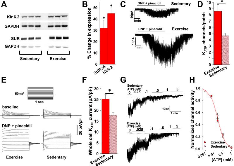
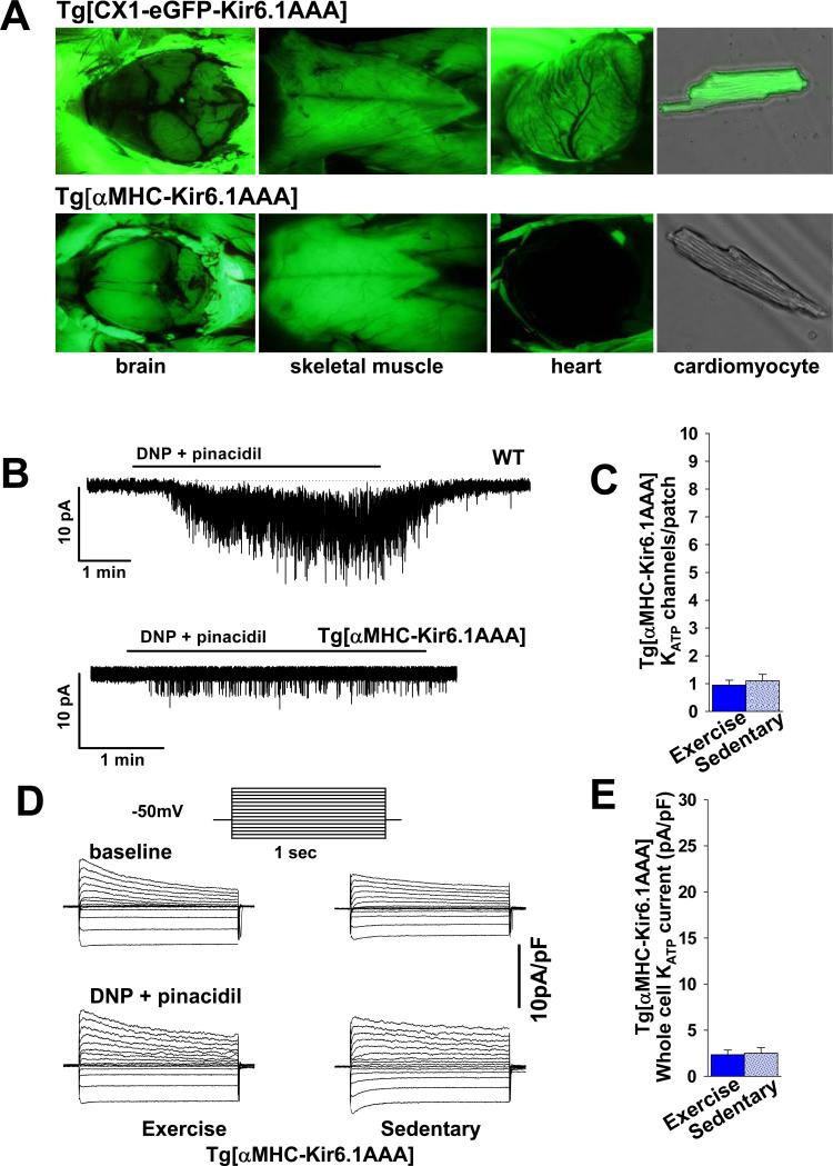
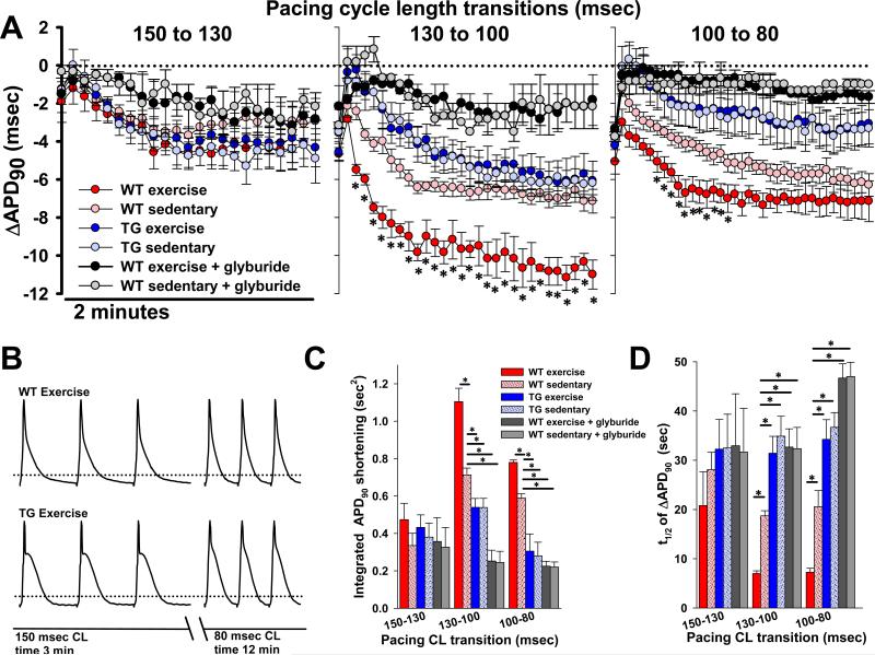
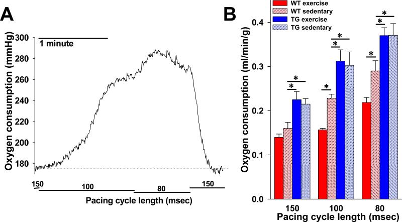
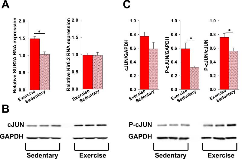
Physical activity is one of the most important determinants of cardiac function. The ability of the heart to increase delivery of oxygen and metabolic fuels relies on an array of adaptive responses necessary to match bodily demand while avoiding exhaustion of cardiac resources. The ATP-sensitive potassium (K(ATP)) channel has the unique ability to adjust cardiac membrane excitability in accordance with ATP and ADP levels, and up-regulation of its expression that occurs in response to exercise could represent a critical element of this adaption. However, the mechanism by which K(ATP) channel expression changes result in a beneficial effect on cardiac excitability and function remains to be established. Here, we demonstrate that an exercise-induced rise in K(ATP) channel expression enhanced the rate and magnitude of action potential shortening in response to heart rate acceleration. This adaptation in membrane excitability promoted significant reduction in cardiac energy consumption under escalating workloads. Genetic disruption of normal K(ATP) channel pore function abolished the exercise-related changes in action potential duration adjustment and caused increased cardiac energy consumption. Thus, an expression-driven enhancement in the K(ATP) channel-dependent membrane response to alterations in cardiac workload represents a previously unrecognized mechanism for adaptation to physical activity and a potential target for cardioprotection.


Figures

Figure 1
Figure 1. Short-term exercise increases cardiac expression of functional KATP channels
A. Representative Western blots of KATP channel subunits Kir6.2 and SUR2A in protein extracts from ventricles of mice under sedentary and exercise conditions. B. Summary statistics indicating increased expression of Kir6.2 and SUR2A subunits over baseline in response to exercise (both n=10 and *p<.05 vs. baseline). C. Representative cell-attached KATP channel recordings in ventricular myocytes isolated from hearts of sedentary and exercised mice. Channel opening is stimulated by 2,4-dinitrophenol (DNP) 200μM and pinacidil 100μM in the perfusate. D. Summary statistics indicating the number of KATP channels/patch detected in cardiomyocytes isolated from exercised (n=22 patches, 5 mice) and sedentary (n=29 patches, 6 mice) animals (*p<.001). The patch pipette tip size was not significantly different between groups (4.71±.31 vs. 4.65±.22 MΩ, respectively, p=NS). E. Representative whole cell KATP current recordings in cardiomyocytes isolated from hearts of exercised and sedentary mice in response to DNP 50μM and pinacidil 100μM in the perfusate. Inset: stimulation pattern. Current is normalized to membrane capacitance (pA/pF). F. Summary statistics of whole cell KATP channel current normalized to cell capacitance (pA/pF) in response to DNP 50μM and pinacidil 100μM for cardiomyocytes isolated from hearts of exercised (n=19 patches, 5 mice) vs. sedentary (n=10 patches, 6 mice) animals (* p<.05). G. Representative inside-out patch recordings of KATP channel activity in isolated cardiomyocytes from hearts of exercised and sedentary mice in response to escalating concentrations of ATP in the perfusate. H. KATP channel activity in membrane patches (inside-out configuration), calculated relative to activity in the absence of ATP, and fitted by the Hill equation (y=[IC50]/([IC50]h + [ATP]h), where h is the Hill coefficient, [ATP] is the ATP concentration, and [IC50] is the half-maximal inhibitory ATP concentration (n=10 exercise and 9 sedentary).
Figure 2
Figure 2. Expression of a dominant negative KATP channel pore-forming subunit results in significant reduction of functional cardiac channels and interference with exercise-induced KATP current up-regulation
A. Representative images of tissues from transgenic mice under UV light. Tg[CX1-eGFP-Kir6.1AAA] mice express eGFP in all cells (top panel). Crossing Tg[CX1-eGFP-Kir6.1AAA] with Tg[αMHC-Cre] generated Tg[αMHC-Kir6.1AAA] progeny with cardiomyocyte specific Kir6.1AAA transgene expression tracked by selective elimination of eGFP fluorescence in the heart only (bottom panel). B. Representative cell-attached KATP channel recordings in response to DNP 200μM and pinacidil 100μM in the perfusate. C. Summary statistics indicating the number of KATP channels/patch in cell-attached recordings following exposure to DNP 200μM and pinacidil 100μM performed in cardiomyocytes isolated from exercised (n=5 mice and 17 patches) vs. sedentary (n=5 mice, 10 patches) Tg[αMHC-Kir6.1AAA] animals (p=NS). The patch pipette tip size was not significantly different between groups (4.62±.09 vs. 4.44±.07 MΩ, respectively, p=NS). D. Representative whole cell KATP channel current recordings in cardiomyocytes isolated from hearts of exercised and sedentary Tg[αMHC-Kir6.1AAA] mice in response to DNP 50μM and pinacidil 100μM in the perfusate. Inset shows stimulation pattern. Current is normalized to cell capacitance (pA/pF). E. Summary statistics of whole cell KATP channel current normalized to cell capacitance (pA/pF) in response to DNP 50μM and pinacidil 100μM for cardiomyocytes isolated from hearts of exercised (n= 3 mice and 11 patches) vs. sedentary (n= 3 mice and 11 patches) Tg[αMHC-Kir6.1AAA] animals (p=NS).
Figure 3
Figure 3. Exercise-induced increase in KATP channel expression translates to enhanced action potential shortening in response to heart rate acceleration
A. Summary plots of changes in monophasic action potential shortening (ΔAPD90) as a function of time following each of three pacing cycle length transitions in isolated, perfused hearts from WT exercised (n=7), WT sedentary (n=8), TG exercised (n=7), and TG sedentary (n=7) mice (*p<.05 for WT exercise vs. WT sedentary). Also graphed are glyburide (10μM) treated hearts from WT exercise (n=3) and WT sedentary mice (n=3). Each graph indicates shortening as compared to the steady state APD90 measured for the previous pacing cycle length. There is no difference in steady state APD90 after 3 minutes of pacing at 150 msec. (59.3±3.5, 57.4±3, 59.6±3.3, 57.4±4.1, 60.3±4.1 and 59.5±3.8 msec, respectively, p=NS). The ΔAPD90 of the first action potential following the transition is graphed, followed by every 40th action potential thereafter for 2 minutes. B. Representative examples of monophasic action potentials recorded from the left ventricular epicardium of isolated, perfused hearts from exercised WT and Tg[αMHC-Kir6.1AAA] mice after 3 minutes of pacing at the displayed cycle length, in accordance with the 12 minute protocol of progressive cycle length shortening. Dotted lines indicate the 90% repolarization level. C. Summary statistics showing the integrated APD90 shortening, defined as the area above the curve of the ΔAPD90 time course (n = same as in Fig. 3A, *p<.05 compared to WT sedentary). D. Summary statistics showing the half time (t1/2) of ΔAPD90 response (n = same as in Fig. 3A, *p<.05 compared to WT sedentary). WT = wild-type, TG = Tg[αMHC-Kir6.1AAA].
Figure 4
Figure 4. Rate of cardiac oxygen consumption in response to heart rate acceleration is reduced in animals with increased KATP channel expression following exercise
A. Representative tracing of raw oxygen consumption data (perfusate oxygen tension – effluent oxygen tension) in response to pacing cycle length transitions in an isolated perfused heart from a WT sedentary animal. B. Summary statistics indicating steady state oxygen consumption rate (V̇O2), normalized to heart weight and coronary flow (see Methods), at each of three pacing cycle lengths in isolated perfused hearts from WT exercise (n=7), WT sedentary (n=7), TG exercise (n=4) and TG sedentary (n=5) mice (*p<.05 compared to WT sedentary). WT = wild-type, TG = Tg[αMHC-Kir6.1AAA].
Figure 5
Figure 5. Increased cardiac KATP channel expression in response to exercise is associated with increased transcription of the SUR2A encoding gene
A. Summary of the relative levels of SUR2A (left bar graph) and Kir6.2 (right bar graph) mRNA in extracts from ventricles isolated from hearts of exercised and sedentary mice (n=9 for each group, *p<.05). B. Representative western blots of cJUN and P-cJUN compared to GAPDH in extracts from ventricles isolated from hearts of exercised and sedentary mice. C. Summary of cJUN and phorphorylated cJUN (P-cJUN) expression relative to GAPDH in extracts from ventricles of hearts from exercised (n=3) and sedentary (n=3) mice (*p<.05 for Pc-JUN and Pc-JUN/cJUN exercise vs. sedentary).

Similar articles

Cited by

References

    1. Guyton AC, Hall JE. Sport physiology. In: Guyton AC, Hall JE, editors. Textbook of medical physiology. Elsevier Saunders; Philadelphia, PA: 2006. pp. 1062–1062-1065.
    1. Aguilar-Bryan L, Bryan J. Molecular biology of adenosine triphosphate-sensitive potassium channels. Endocr Rev. 1999;20:101–35. - PubMed
    1. Noma A. ATP-regulated K+ channels in cardiac muscle. Nature. 1983;305:147–8. - PubMed
    1. Seino S, Miki T. Physiological and pathophysiological roles of ATP-sensitive K+ channels. Prog Biophys Mol Biol. 2003;81:133–76. - PubMed
    1. Flagg TP, Enkvetchakul D, Koster JC, Nichols CG. Muscle KATP channels: recent insights to energy sensing and myoprotection. Physiol Rev. 2010;90:799–829. - PMC - PubMed

Publication types