

Intraprocedure visualization of the esophagus using interventional C-arm CT as guidance for left atrial radiofrequency ablation
- PMID: 21440465
- PMCID: PMC3115455
- DOI: 10.1016/j.acra.2011.01.023
Intraprocedure visualization of the esophagus using interventional C-arm CT as guidance for left atrial radiofrequency ablation
Abstract
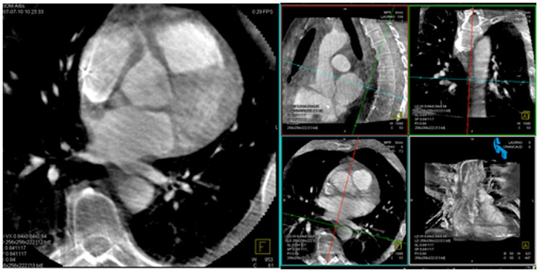
Rationale and objectives: During radiofrequency catheter ablation for atrial fibrillation, the esophagus is at risk for thermal injury. In this study, C-arm computed tomography (CT) was compared to clinical CT, without the administration of oral contrast, to visualize the esophagus and its relationship to the left atrium and the ostia of the pulmonary veins (PVs) during the radiofrequency ablation procedure.
Materials and methods: Sixteen subjects underwent both cardiac clinical CT and C-arm CT. Computed tomographic scans were performed on a multidetector scanner using a standard electrocardiographically gated protocol. C-arm computed tomographic scans were obtained using either a multisweep protocol with retrospective electrocardiographic gating or a non-gated single-sweep protocol. C-arm and clinical computed tomographic scans were analyzed in a random order and then compared for the following criteria: (1) visualization of the esophagus (yes or no), (2) relationship of esophageal position to the four PVs, and (3) direct contact or absence of a fat pad between the esophagus and the PV antrum.
Results: The esophagus was identified in all C-arm and clinical computed tomographic scans. In four cases, orthogonal planes were needed on C-arm CT (inferior PV level). In six patients, the esophageal location on C-arm CT was different from that on CT. Direct contact was reported in 19 of 64 of the segments (30%) examined on CT and in 26 of 64 (41%) on C-arm CT. In five of 64 segments (8%), C-arm CT overestimated a direct contact of the esophagus to the left atrium.
Conclusions: C-arm computed tomographic image quality without the administration of oral contrast agents was shown to be sufficient for visualization of the esophagus location during a radiofrequency catheter ablation procedure for atrial fibrillation.
Copyright © 2011 AUR. Published by Elsevier Inc. All rights reserved.
Figures


Similar articles
-
Esophagus imaging for catheter ablation of atrial fibrillation: comparison of two methods with showing of esophageal movement.J Interv Card Electrophysiol. 2009 Dec;26(3):159-64. doi: 10.1007/s10840-009-9434-3. Epub 2009 Sep 10. J Interv Card Electrophysiol. 2009. PMID: 19757002 Clinical Trial.
-
Esophageal positions relative to the left atrium; data from 293 patients before catheter ablation of atrial fibrillation.Indian Heart J. 2018 Jan-Feb;70(1):37-44. doi: 10.1016/j.ihj.2017.06.013. Epub 2017 Jun 29. Indian Heart J. 2018. PMID: 29455785 Free PMC article.
-
Three-dimensional esophagus reconstruction and monitoring during ablation of atrial fibrillation: combination of two imaging techniques.Int J Cardiol. 2013 Oct 3;168(3):2364-8. doi: 10.1016/j.ijcard.2013.01.026. Epub 2013 Feb 14. Int J Cardiol. 2013. PMID: 23416012
-
[Three-dimensional reconstruction and remote navigation for catheter-guided atrial fibrillation ablation. Does it influence procedural outcomes?].Clin Res Cardiol Suppl. 2011 May;6:73-7. doi: 10.1007/s11789-011-0028-0. Clin Res Cardiol Suppl. 2011. PMID: 22528181 Review. German.
-
The role of computed tomography and magnetic resonance imaging in ablation procedures for treatment of atrial fibrillation.Semin Ultrasound CT MR. 2009 Apr;30(2):125-56. doi: 10.1053/j.sult.2008.10.015. Semin Ultrasound CT MR. 2009. PMID: 19358443 Review.
Cited by
-
The use of intracardiac echocardiography catheters in endocardial ablation of cardiac arrhythmia: Meta-analysis of efficiency, effectiveness, and safety outcomes.J Cardiovasc Electrophysiol. 2020 Mar;31(3):664-673. doi: 10.1111/jce.14367. Epub 2020 Jan 30. J Cardiovasc Electrophysiol. 2020. PMID: 31976603 Free PMC article.
References
-
- Haissaguerre M, Jais P, Shah DC, et al. Spontaneous initiation of atrial fibrillation by ectopic beats originating in the pulmonary veins. N Engl J Med. 1998;339(10):659–666. - PubMed
-
- Tsao HM, Wu MH, Higa S, et al. Anatomic relationship of the esophagus and left atrium: implication for catheter ablation of atrial fibrillation. Chest. 2005;128(4):2581–2587. - PubMed
-
- Jais P, Haissaguerre M, Shah DC, Chouairi S, Clementy J. Regional disparities of endocardial atrial activation in paroxysmal atrial fibrillation. Pacing Clin Electrophysiol. 1996;19(11 Pt 2):1998–2003. - PubMed
-
- Wongcharoen W, Tsao HM, Wu MH, et al. Morphologic characteristics of the left atrial appendage, roof, and septum: implications for the ablation of atrial fibrillation. J Cardiovasc Electrophysiol. 2006;17(9):951–956. - PubMed
-
- Chiam PT, Ruiz CE. Percutaneous transcatheter left atrial appendage exclusion in atrial fibrillation. J Invasive Cardiol. 2008;20(4):E109–E113. - PubMed
MeSH terms
Grants and funding
LinkOut - more resources
Full Text Sources
Medical
Research Materials
