Review
. 2011 Jun;27(6):254-63.
doi: 10.1016/j.pt.2011.02.006. Epub 2011 Mar 26.

Tissue destruction and invasion by Entamoeba histolytica

Affiliations
Review

Tissue destruction and invasion by Entamoeba histolytica

Katherine S Ralston et al. Trends Parasitol. 2011 Jun.

Abstract

Entamoeba histolytica is the causative agent of amebiasis, a disease that is a major source of morbidity and mortality in the developing world. The potent cytotoxic activity of the parasite appears to underlie disease pathogenesis, although the mechanism is unknown. Recently, progress has been made in determining that the parasite activates apoptosis in target cells and some putative effectors have been identified. Recent studies have also begun to unravel the host genetic determinants that influence infection outcome. Thus, we are beginning to get a clearer picture of how this parasite manages to infect, invade and ultimately inflict devastating tissue destruction.


Figures

Figure 1
Figure 1. Tissue destruction associated with E. histolytica infection
(a) An example of amebic colitis, showing the presence of multiple ulcers. (b) A side-view of the classical flask-shaped ulcer seen in amebic colitis. (c) E. histolytica trophozoites (arrows) taken from an ulcer, showing the presence of numerous ingested red blood cells. Panels (a–b) are reproduced from Ref. [83], with permission.
Figure 2
Figure 2. Leptin signaling influences tissue destruction during amebic infection
(a) Chart illustrating how leptin receptor polymorphisms at position 223 influence susceptibility to infection. Shown is Kaplan-Meier survival free of E. histolytica infection of children (n=185) with Gln/Gln (QQ), Gln/Arg (QR) or Arg/Arg (RR) genotypes. (b) Mice carrying one or two copies of the arginine allele at the 223 codon were more susceptible to intestinal infection with E. histolytica than those homozygous for the glutamine (QQ) allele (QQ versus QR p = 0.009, QQ versus RR p = 0.001 by Chi-square test). (c) Mice deficient for leptin (ob/ob) or the functional leptin receptor (db/db) exhibit dramatic destruction of the mucosal epithelium upon intracecal E. histolytica infection. Shown is the normal epithelium in an infected wild-type B6 mouse for comparison. Panels (a–b) are reproduced from Ref. [12] and panel (c) is reproduced from Ref. [13], with permission.
Figure 3
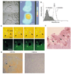
Figure 3. E. histolytica induces calcium influx, tyrosine dephosphorylation and caspase-3 dependent host cell death
(a–c) Time series of Fura-2-loaded CHO cells before contact (top panels) and 30 s after (bottom panels) contact with an E. histolytica (Eh) trophozoite. (a) Phase images and (b) digitized R340/380 images are shown. Bar, 10 µm (c) Color bar indicating intensity from background (dark blue) to a maximal R340/380 (red) of Fura-2 fluorescence. (d) FACS analysis of E. histolytica-induced protein dephosphorylation in Jurkat cells. Cells were incubated with or without E. histolytica, fixed, permeabilized and stained with FITC-pTyr-MAb PT-66. The data express the fluorescence intensities from Jurkat cells; cells in medium alone are shown with the dotted line; cells that were incubated with amebae are shown with the continuous line and shaded area. (e) E. histolytica activates caspase-3 in a contact-dependent manner in vitro. A time series of E. histolytica and Jurkat T cells pre-loaded with the fluorescent caspase-3 substrate rhodamine-G1D2-rhodamine (PhiPhiLux) is shown. Images at 0, 8, 16 and 20 minutes are shown. One Jurkat cell is already positive for active caspase-3 (showing green fluorescence) at time zero. The other Jurkat cells (arrows) show active caspase-3 only following contact with the trophozoites. (f) TUNEL staining in a mouse model of amebic colitis. TUNEL positive apoptotic cells surround E. histolytica trophozoites (arrows) in the infected caecum. (g) Pre-treatment with the pan caspase inhibitor zVAD-fmk reduces the formation of liver abscesses in a mouse model. TUNEL staining of liver sections is shown from mice that were pre-treated with zVAD-fmk or left untreated prior to amebic infection. The tissue has a normal appearance and is mostly TUNEL negative in the treated example, while in the untreated sample the tissue is TUNEL positive, appears dead and has numerous detectable E. histolytica trophozoites (arrows). Panels (a–c) are reproduced from Ref. [33], panel (d) is reproduced from Ref. [36], panels (e) and (f) are reproduced from Ref. [41], and panel (g) is reproduced from Ref. [42], with permission.
Figure 4
Figure 4. Model for the mechanism of cytotoxicity
E. histolytica (blue) is shown adhered to the targeted host cell (tan) via the action of the parasite Gal/GalNAc lectin (green rectangles). Parasite acidic vesicles (purple circles) that are hypothesized to play a role in cytotoxicity are shown; it is possible that their contents are released at a localized synapse between host and parasite. It is unknown whether any parasite effector molecules are transferred to the target cell, but candidates include KERP1 (pink rectangles), the amoebapores and other members of the SAPLIP family (orange donuts). Note that there are additional proteins besides the Gal/GalNAc lectin that influence host cell binding (orange rectangles, turquoise rectangles) such as TMKB1-9 and the EhSTIRPs. The Gal/GalNAc lectin may itself contribute to the cytotoxic mechanism, either by triggering the initiation of the cytotoxic program and/or by triggering calcium influx. Note also that the intermediate and light chains of the lectin (igl, lgl) may play some role in host cell killing. A dramatic influx of extracellular calcium (Ca2+, pink circles) is required for target cell killing; the mechanism for calcium entry is unknown, though it may involve the amoebapores and other members of the SAPLIP family. Calcium influx appears to activate calpain, which proteolytically activates PTP1b, that in turn results in tyrosine dephosphorylation (tyrosine de-P). However, PTP1b is unlikely to be the only PTPase responsible for the observed tyrosine dephosphorylation. Ultimately, caspase-3 becomes activated (caspase-3*) and the target cell succumbs to apoptotic death; however the specific upstream signals that activate caspase-3 are unknown.

Similar articles

Cited by

References

    1. WHO/PAHO/UNESCO report. A consultation with experts on amoebiasis. Mexico City, Mexico 28–29 January, 1997. Epidemiol Bull. 1997;18:13–14. - PubMed
    1. Mondal D, et al. Entamoeba histolytica-associated diarrheal illness is negatively associated with the growth of preschool children: evidence from a prospective study. Trans R Soc Trop Med Hyg. 2006;100:1032–1038. - PubMed
    1. Petri WA, Jr, et al. The bittersweet interface of parasite and host: lectin-carbohydrate interactions during human invasion by the parasite Entamoeba histolytica. Annu Rev Microbiol. 2002;56:39–64. - PubMed
    1. Petri WA., Jr Therapy of intestinal protozoa. Trends Parasitol. 2003;19:523–526. - PubMed
    1. Pritt BS, Clark CG. Amebiasis. Mayo Clin Proc. 2008;83:1154–1159. quiz 1159–1160. - PubMed

Publication types

MeSH terms