. 2011 Jul;141(1):378-88, 388.e1-4.
doi: 10.1053/j.gastro.2011.03.044. Epub 2011 Mar 24.

A mouse model of cholestasis-associated cholangiocarcinoma and transcription factors involved in progression

Affiliations

A mouse model of cholestasis-associated cholangiocarcinoma and transcription factors involved in progression

Heping Yang et al. Gastroenterology. 2011 Jul.

Abstract

Background & aims: Cholestasis contributes to hepatocellular injury and promotes liver carcinogenesis. We created a mouse model of chronic cholestasis to study its effects on progression of cholangiocarcinoma and the oncogenes involved.

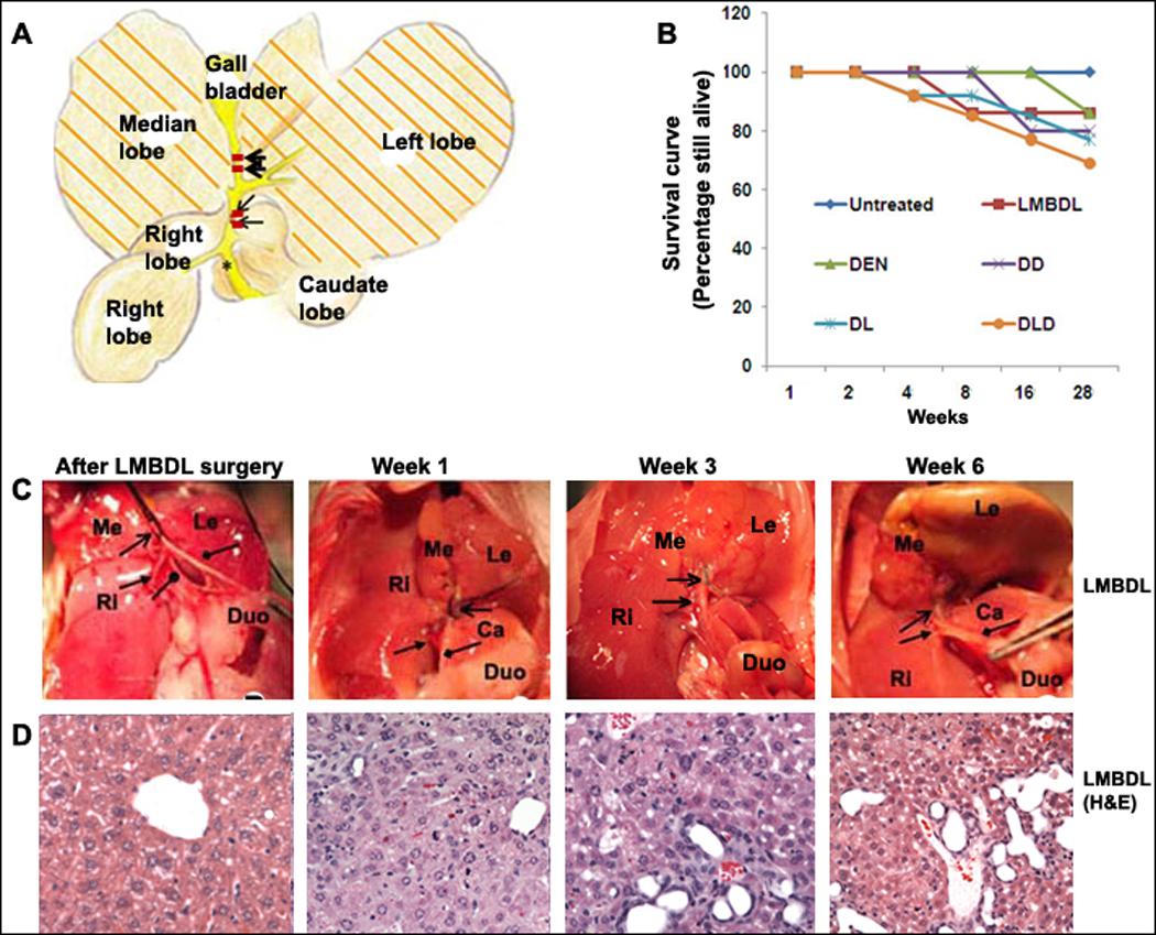
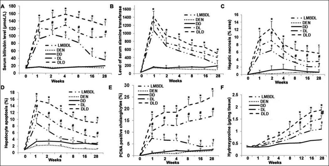
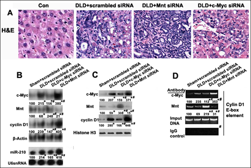
Methods: To induce chronic cholestasis, Balb/c mice were given 2 weekly intraperitoneal injections of diethylnitrosamine (DEN); 2 weeks later, some mice also received left and median bile duct ligation (LMBDL) and, then 1 week later, were fed DEN, in corn oil, weekly by oral gavage (DLD). Liver samples were analyzed by immunohistochemical and biochemical assays; expression of Mnt and c-Myc was reduced by injection of small inhibitor RNAs.

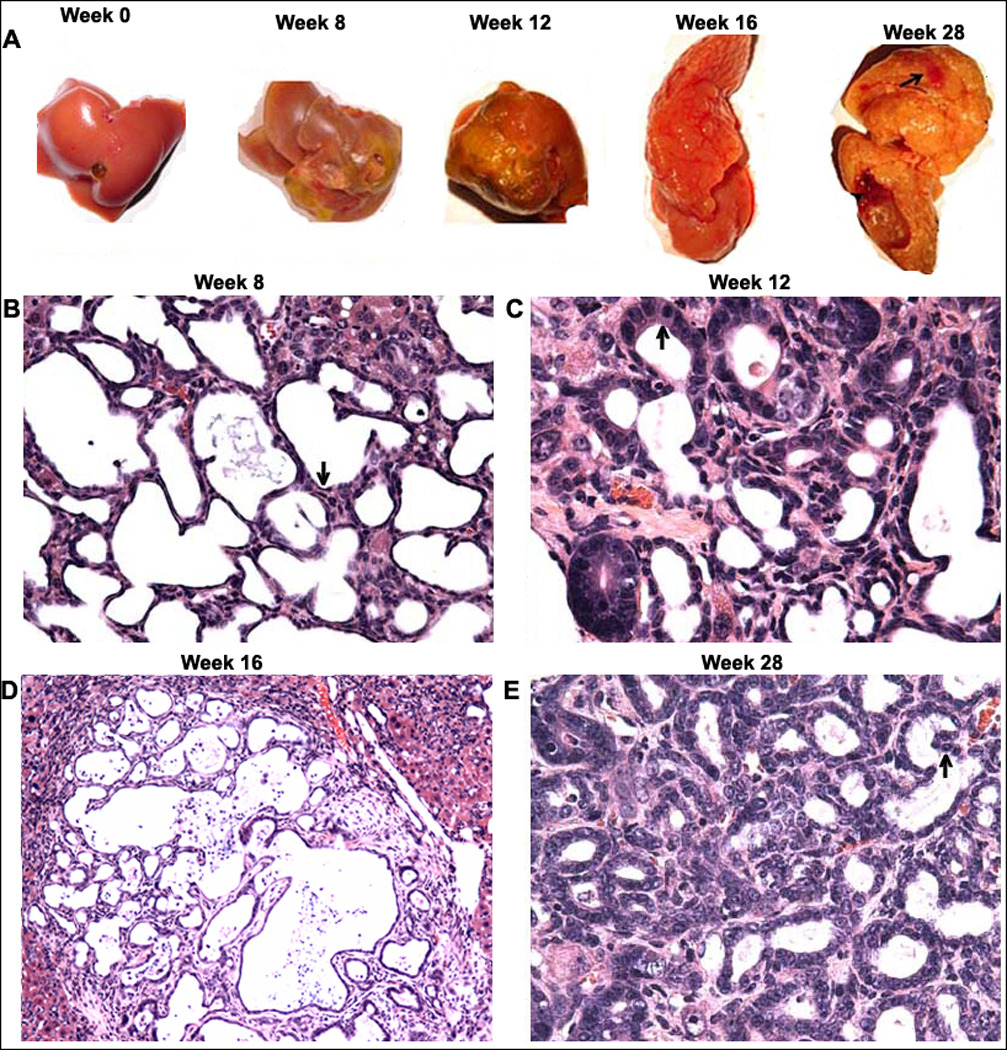
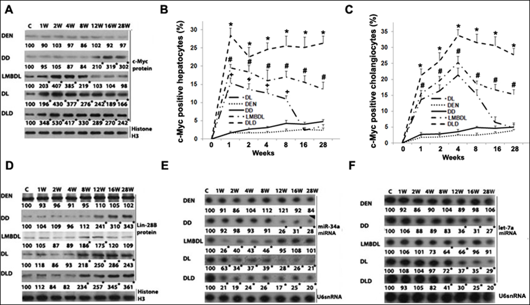
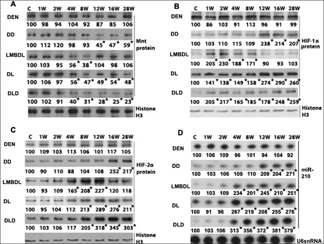
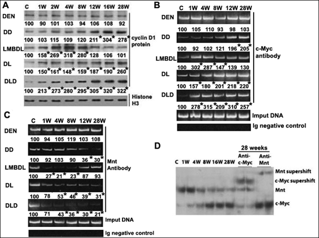
Results: Chronic cholestasis was induced by DLD and accelerated progression of cholangiocarcinoma, compared with mice given only DEN. Cystic hyperplasias, cystic atypical hyperplasias, cholangiomas, and cholangiocarcinoma developed in the DLD group at weeks 8, 12, 16, and 28, respectively. LMBDL repressed expression of microRNA (miR)-34a and let-7a, up-regulating Lin-28B, hypoxia-inducible factor (HIF)-1α, HIF-2α, and miR-210. Up-regulation of Lin-28B might inhibit let-7a, which is associated with development of cystic hyperplasias, cystic atypical hyperplasias, cholangiomas, and cholangiocarcinoma. Knockdown of c-Myc reduced progression of cholangiocarcinoma, whereas knockdown of Mnt accelerated its progression. Down-regulation of miR-34a expression might up-regulate c-Myc. The up-regulation of miR-210 via HIF-2α was involved in down-regulation of Mnt. Activation of the miR-34a-c-Myc and HIF-2α-miR-210-Mnt pathways caused c-Myc to bind the E-box element of cyclin D1, instead of Mnt, resulting in cyclin D1 up-regulation.

Conclusions: DLD induction of chronic cholestasis accelerated progression of cholangiocarcinoma, which is mediated by down-regulation of miR-34a, up-regulation miR-210, and replacement of Mnt by c-Myc in binding to cyclin D1.


Conflict of interest statement

Potential conflict of interest: Nothing to report.

Figures

Figure 1
Figure 1. The effect of LMBDL on the hepatobiliary system and survival rate
A) A diagram of the ligation position for LMBDL. B) Survival curve of mice with different treatments. Mice treated with LMBDL (6 of 7), DEN (6 of 7), DD (8 of 10), DL (10 of 13) and DLD (10 of 14) exhibited a high survival rate at 28 weeks. C) The hepatobiliary system after 1, 3 and 6 weeks. Arrow end type (↑) =position of left and median bile duct ligation, triangle arrow end type= right hepatic bile duct, rhombus arrow end type= common bile duct, circle arrow end type=caudate hepatic bile duct. D) H&E staining of the livers from the LMBDL mice after the indicated time points (100X).
Fig. 2
Fig. 2. Assessment of liver damage and histopathological features in the ligated lobes of the treated mice
Total serum was extracted from the treated groups to assess A) total bilirubin and B) ALT levels. C) Hepatic necrosis was determined by H&E staining. D) Hepatocyte apoptosis was identified by TUNEL staining. E) Cholangiocyte proliferation was evaluated by PCNA staining. F) Hydroxyproline content was done to look at fibrosis. Five liver tissue samples for each group were used for the above assays. *p<0.05 DLD vs all other groups, #p<0.05 DL vs LMBDL, p<0.01 LMBDL vs DEN and DD groups.
Fig. 3
Fig. 3. Time course of progressive liver injury and morphological analysis of CCA in the ligated lobes of the DLD group
A) Representative livers showing fibrosis (week 8), cirrhosis (week 12), cholangiomas (week 16) and CCA (week 28- denoted by arrow). H&E staining was done to assess liver morphology at the various time points. B) A week 8 liver showing multifocal cystic hyperplasia of the intrahepatic bile ducts and multifocal cysts lined by flattened epithelium (arrow) (200×); C) A week 12 liver showing cystic atypical hyperplasia of the intrahepatic bile ducts and multifocal cysts lined by homogenous epithelium with elongated nuclei (arrow) (200×); D) A week 16 liver showing cholangioma formation and ductular adenoma formation (100×); and E) A week 28 liver showing CCA formation (200×).
Fig. 4
Fig. 4. Time-course of c-Myc expression in mice treated with DEN, DD, LMBDL, DL and DLD
A) Western blot assays of c-Myc protein expression. c-Myc immunohistochemistry assays showing the percentage of c-Myc-positive staining in B) hepatocytes and C) cholangiocytes. D) Western blot assays for Lin-28B. Northern blot assays for E) miR-34a and F) let-7a. *p<0.01 DLD vs all other groups, #p<0.01 DL vs LMBDL, †p<0.01 LMBDL vs DEN and DD groups, and *p<0.01 vs control.
Fig. 5
Fig. 5. Time- course of HIF-1α, HIF-2α, Mnt and miR-210 expression
Western blot analysis of the treated groups from day 0 to 28 weeks looking at A) Mnt, B) HIF-1α and C) HIF-2α. D) Northern blot analysis of miR-210 in the treated groups from day 0 to 28 weeks. *p<0.01 vs control.
Fig. 6
Fig. 6. Expression and binding activities of c-Myc and Mnt on the E-box in the cyclin D1 promoter
A) Western blot analysis of cyclin D1 in mice treated with DEN, DD, LMBDL, DL and DLD from day 0 to 28 weeks. *p<0.01 vs control. ChiP assays of the E-box element in cyclin D1 promoter was done using antibodies to either B) c-Myc or C) Mnt. D) EMSA of cyclin D1 promoter (−425 to −446 of the mouse E-box region) and supershift analysis with c-Myc or Mnt antiboby. *p<0.01 vs control.
Fig. 7
Fig. 7. Effect of c-Myc or Mnt siRNA on liver morphology, gene expression and binding activity to the E-box element of cyclin D1 promoter in DLD mice
siRNA knockdown of either c-Myc or Mnt affecting changes to A) Liver morphology (H&E staining, 200X), B) c-Myc, Mnt, cyclin D1 and miR-210 mRNA expression, C) c-Myc, Mnt and cyclin D1 protein expression, and D) Binding activity of c-Myc, Mnt and in the E-box region of cyclin D1 promoter by ChiP analysis. *p<0.01 vs Sham+scrambled siRNA, #<0.01 DLD+c-Myc siRNA or Mnt siRNA vs DLD+scrambled siRNA.

Comment in

Similar articles

Cited by

References

    1. Guglielmi A, Ruzzenente A, Campagnaro T, et al. Does intrahepatic cholangiocarcinoma have better prognosis compared to perihilar cholangiocarcinoma? J Surg Oncol. 2010;101:111–115. - PubMed
    1. Lazaridis KN, Gores GJ. Cholangiocarcinoma. Gastroenterology. 2005;128:1655–1667. - PubMed
    1. Welzel TM, Graubard BI, El-Serag HB, et al. Risk factors for intrahepatic and extrahepatic cholangiocarcinoma in the United States: a population-based case-control study. Clin Gastroenterol Hepatol. 2007;5:1221–1228. - PMC - PubMed
    1. Sirica AE, Dumur CI, Campbell DJ, Almenara JA, Ogunwobi OO, Dewitt JL. Intrahepatic cholangiocarcinoma progression: prognostic factors and basic mechanisms. Clin Gastroenterol Hepatol. 2009;7(11 Suppl):S68–S78. - PMC - PubMed
    1. Yang H, Li TW, Ko KS, Xia M, Lu SC. Switch from Mnt-Max to Myc-Max induces p53 and cyclin D1 expression and apoptosis during cholestasis in mouse and human hepatocytes. Hepatology. 2009;49:860–870. - PMC - PubMed

Publication types

MeSH terms

Substances