. 2011 May;10(5):662-71.
doi: 10.1128/EC.00221-10. Epub 2011 Mar 25.

Uncovering a role for the tail of the Dictyostelium discoideum SadA protein in cell-substrate adhesion

Affiliations

Uncovering a role for the tail of the Dictyostelium discoideum SadA protein in cell-substrate adhesion

Anthony S Kowal et al. Eukaryot Cell. 2011 May.

Abstract

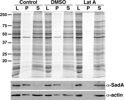
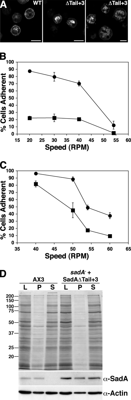
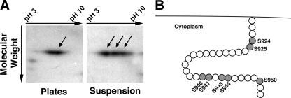
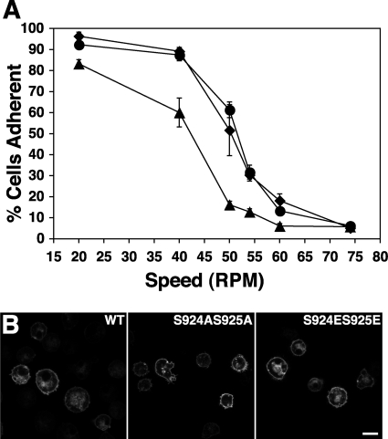
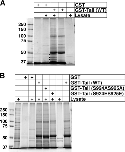
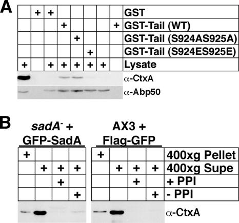
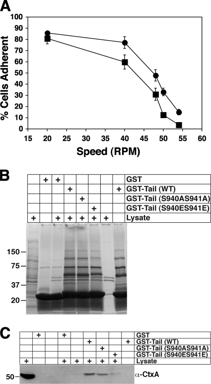
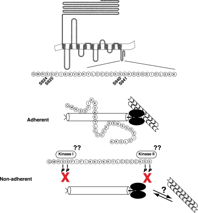
Previous work from our laboratory showed that the Dictyostelium discoideum SadA protein plays a central role in cell-substrate adhesion. SadA null cells exhibit a loss of adhesion, a disrupted actin cytoskeleton, and a cytokinesis defect. How SadA mediates these phenotypes is unknown. This work addresses the mechanism of SadA function, demonstrating an important role for the C-terminal cytoplasmic tail in SadA function. We found that a SadA tailless mutant was unable to rescue the sadA adhesion deficiency, and overexpression of the SadA tail domain reduced adhesion in wild-type cells. We also show that SadA is closely associated with the actin cytoskeleton. Mutagenesis studies suggested that four serine residues in the tail, S924/S925 and S940/S941, may regulate association of SadA with the actin cytoskeleton. Glutathione S-transferase pull-down assays identified at least one likely interaction partner of the SadA tail, cortexillin I, a known actin bundling protein. Thus, our data demonstrate an important role for the carboxy-terminal cytoplasmic tail in SadA function and strongly suggest that a phosphorylation event in this tail regulates an interaction with cortexillin I. Based on our data, we propose a model for the function of SadA.


Figures

Fig. 1.
Fig. 1.
SadA associates with the F-actin cytoskeleton. AX3 cells grown under adherent conditions received fresh medium (control) or were treated with DMSO or 5 μM latrunculin A (LatA). Cells lysed with 1% Triton X-100 (L) were subjected to low-speed centrifugation, divided into pellet (P) and supernatant (S) samples, and fractionated by SDS-PAGE followed by Western blotting. Membranes were probed with a polyclonal SadA antibody followed by a monoclonal anti-actin antibody. In control and DMSO-treated cells, SadA partitioned into the Triton-insoluble pellet, and actin was divided between the pellet and supernatant fractions. When cells were treated with latrunculin A, both SadA and actin were observed to fractionate completely into the Triton X-100-soluble fraction.
Fig. 2.
Fig. 2.
Evidence for functional significance of the SadA carboxy-terminal tail. (A) Micrographs of sadA null cells expressing either a wild-type SadA or a SadA carboxy-terminal deletion construct (SadA ΔTail+3-GFP construct) imaged with a confocal microscope. Increased levels of GFP signal were generally observed in cytoplasmic intracellular vesicles compared to the plasma membrane compartment. The right panel is a micrograph of cells imaged using the agar overlay technique, and the left panel shows cells without overlay. Bar, 10 μm. (B) Adhesion maintenance assay of AX3 wild-type and sadA null cells expressing the SadA ΔTail+3-GFP construct. Whereas AX3 cells (•) exhibited a sheer force-dependent decrease in substrate adhesion, the SadA ΔTail+3-GFP-expressing cells (▪) had minimal adhesion at all speeds tested. (C) Adhesion maintenance of AX3 cells (•) and AX3 cells overexpressing the SadA carboxy-terminal tail (▪). AX3 cells overexpressing the SadA tail were less resistant to increased sheer stress than AX3 wild-type cells. Error bars represent the standard deviations from triplicate or quadruplicate samples. (D) Triton solubility assay of AX3 and sadA plus pTXSadAΔtail+3-GFP. The experiment was conducted as described for Fig. 1, but only under control (untreated) conditions. Whereas wild-type SadA partitioned primarily into the Triton X-100-insoluble fraction, SadAΔTail+3 partitioned, almost equally, between the insoluble and soluble fractions.
Fig. 3.
Fig. 3.
2D and bioinformatic analysis of SadA. (A) AX3 wild-type cells were grown under adherent conditions (plates) or under nonadherent conditions (suspension). Cell lysates were subjected to 2D gel analysis followed by Western blotting with a SadA antibody. Under adherent conditions, only one or two SadA reactive spots were seen. However, when grown in suspension, SadA reactivity was observed in a trail, consisting of three to four distinct spots. This trail may represent posttranslational modifications in the tail (i.e., phosphorylation) resulting in charge- induced isoforms. (B) Cartoon depiction of the cytoplasmic SadA carboxy-terminal tail. Shaded residues highlight the seven serine residues, which are putative sites of phosphorylation in this region. There are no threonines and only one tyrosine residue at position 933 in the tail region.
Fig. 4.
Fig. 4.
Functional and morphological effects of SadA S924 S925 mutations. (A) Adhesion strength of sadA null cells expressing either SadA GFP wild-type protein (•), SadA S924AS925A GFP (♦), or SadA S924ES925E GFP (▴) as measured in an adhesion maintenance assay. Whereas the AX3 and SadA S924AS925A GFP cells exhibited similar sheer force-dependent decreases in adhesion, the SadA S924ES925E mutant was less resistant to the same increases in sheer force. (B) Representative micrographs of confocal sections through sadA null cells expressing the same constructs as used in the adhesion maintenance assay. The sadA-GFP constructs were all capable of localizing to the plasma membrane. Bar, 10 μm.
Fig. 5.
Fig. 5.
Identifying SadA tail-interacting proteins. (A) AX3 wild-type lysates were incubated with glutathione-Sepharose beads prebound with either GST (as a negative control) or with a GST-SadA tail fusion protein (wild-type sequence). GST and GST-tail lanes represent beads treated as described for experimental samples but without added lysate, to identify any nonspecific bands from the purified protein sample. The lysate lane represents beads treated as for the experimental samples but without any added bait protein, to observe proteins that nonspecifically bound to the beads. Three of the unique bands were excised from the gel (marked with black dots) and submitted for protein identification via mass spectrometry. (B) GST pull-down assay using GST-tail (WT), GST-tail (S924AS925A), or GST-tail (S924ES925E) fusion proteins as bait. GST, GST-tail, and the far right lysate samples were obtained as described for panel A. The lysate lane next to the molecular weight marker lane represents 1% of the amount of lysate used for pull-down assays.
Fig. 6.
Fig. 6.
Testing the interaction between cortexillin I and SadA. (A) GST pull-down assay using GST-SadA tail (WT), GST-SadA tail (S924AS925A), or GST-SadA tail (S924ES925E) as bait. GST pull-down assays were carried out as described for Fig. 5, except that samples were treated with latrunculin A both before and subsequent to lysis to disrupt F-actin. The left-most lysate lane represents 1% of lysate, used as input into the pull-down reaction. The right-most lysate lane represents a mock pull-down sample (input lysate that was reacted with baitless beads). Subsequent to the pull-down assays, samples were fractionated on 10% SDS-PAGE gels, transferred to PVDF membranes, probed with an anti-cortexillin I antibody (241-438-1), stripped, and reprobed with an anti-Abp50 antibody. (B) GFP-trap pull-down assay. Prior to the pull-down assay, cells were subjected to Triton X-100 treatment as described for the solubility assays and divided between soluble and insoluble fractions. The 400 × g supernant (soluble) fraction from either sadA cells expressing a GFP-SadA fusion protein (treated with latrunculin A) or from AX3 cells expressing a Flag-GFP fusion protein was applied to GFP-Trap beads and allowed to react. Resultant pull-down samples were subjected to SDS-PAGE, followed by transfer to PVDF. The membrane was probed with the anti-cortexillin I antibody.
Fig. 7.
Fig. 7.
Functional and biochemical effects of SadA S940 S941 mutations. (A) sadA null cells expressing either SadA S940AS941A (•) or SadA S940ES941E (▪) were evaluated in an adhesion maintenance assay, as described for Fig. 4A. The S→E phospho-mimetic mutant exhibited less resistance to sheer force than the S→A non-phospho-mimetic mutant. (B) GST pull-down assays, carried out as described for Fig. 5B. (C) GST pull-down assay, as conducted for Fig. 6A, with anti-cortexillin I antibody as the probe.
Fig. 8.
Fig. 8.
Proposed model for the SadA-cortexillin I interaction. Based on our current data, we propose the following model for the SadA tail-cortexillin I (CtxA) interaction. Under adherent conditions, an interaction occurs between the carboxy-terminal tail of SadA via the S924/5 and S940/1 sites and presumably the coiled-coil region of CtxA. This results in a linkage between SadA and the subcortical actin cytoskeleton. Under nonadherent conditions, or during cellular motility, perhaps, an unknown kinase(s) would phosphorylate sites in the SadA tail, resulting in release of CtxA from the SadA tail. This would temporarily disrupt the link between SadA and the actin cytoskeleton. During the processes of cell motility and phagocytosis, the cell must have a dynamic linkage between the actin cytoskeleton and membrane receptor in order to generate the forces necessary for the cell to move or to engulf a particle, respectively. The interaction between SadA and CtxA in our proposed model might be one mechanism that Dictyostelium has developed to perform these tasks.

Similar articles

Cited by

References

    1. Alibaud L., Cosson P., Benghezal M. 2003. Dictyostelium discoideum transformation by oscillating electric field electroporation. Biotechniques 35:78–80 - PubMed
    1. Appella E., Weber I. T., Blasi F. 1988. Structure and function of epidermal growth factor-like regions in proteins. FEBS Lett. 231:1–4 - PubMed
    1. Baldauf S. L. 2003. The deep roots of eukaryotes. Science 300:1703–1706 - PubMed
    1. Balla S., et al. 2006. Minimotif Miner: a tool for investigating protein function. Nat. Methods 3:175–177 - PubMed
    1. Blom N., Gammeltoft S., Brunak S. 1999. Sequence and structure-based prediction of eukaryotic protein phosphorylation sites. J. Mol. Biol. 294:1351–1362 - PubMed

LinkOut - more resources