. 2011 Mar;23(3):1047-60.
doi: 10.1105/tpc.111.083352. Epub 2011 Mar 25.

The CHD3 chromatin remodeler PICKLE and polycomb group proteins antagonistically regulate meristem activity in the Arabidopsis root

Affiliations

The CHD3 chromatin remodeler PICKLE and polycomb group proteins antagonistically regulate meristem activity in the Arabidopsis root

Ernst Aichinger et al. Plant Cell. 2011 Mar.

Abstract

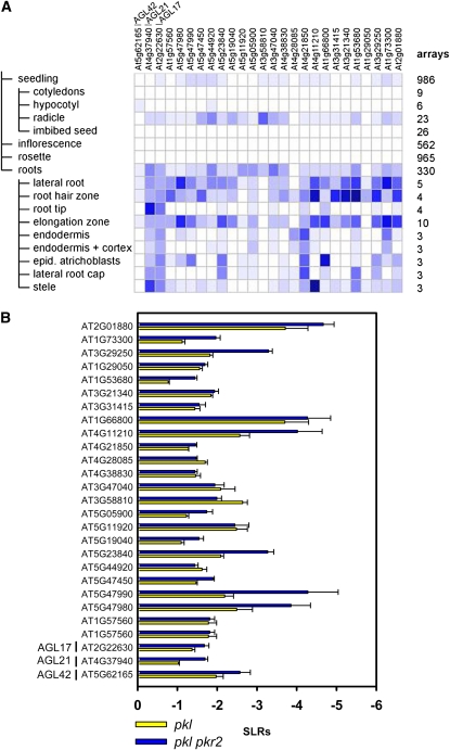
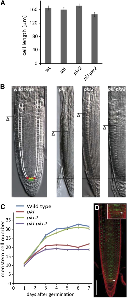
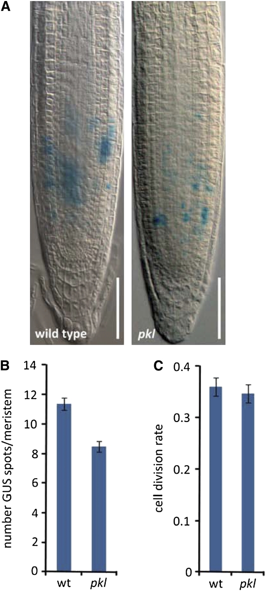
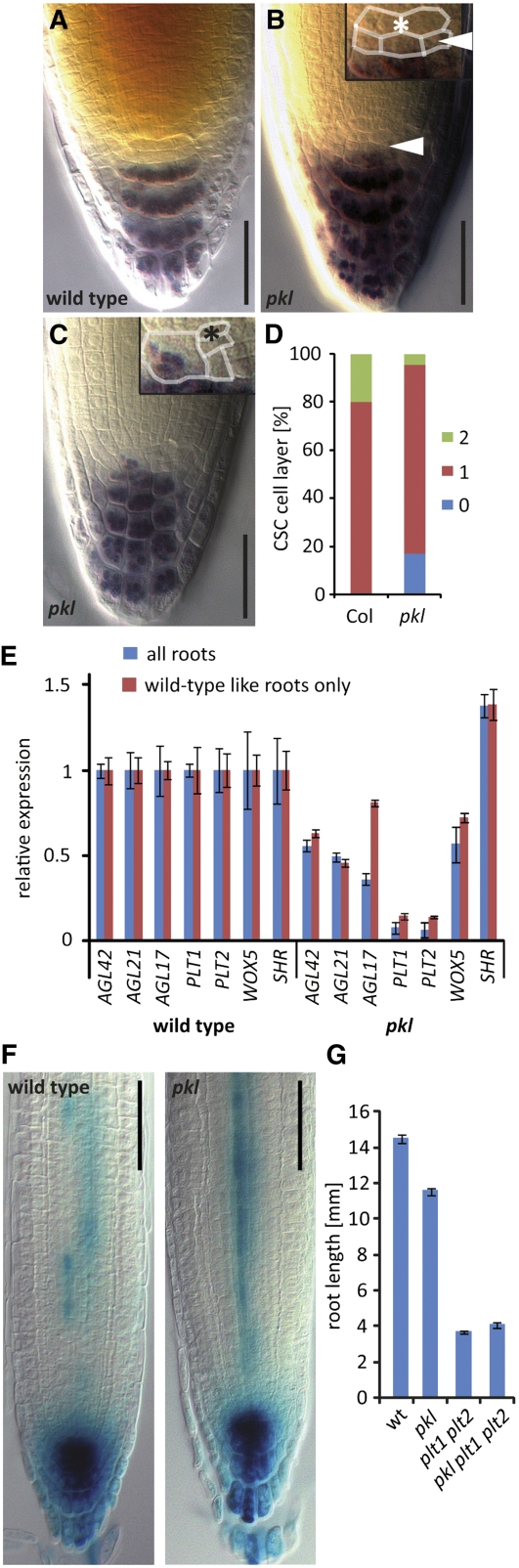
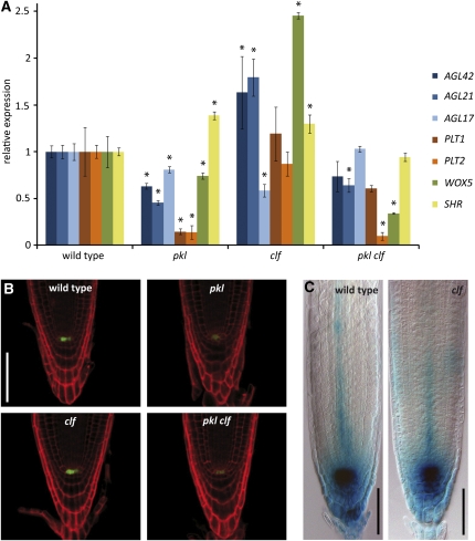
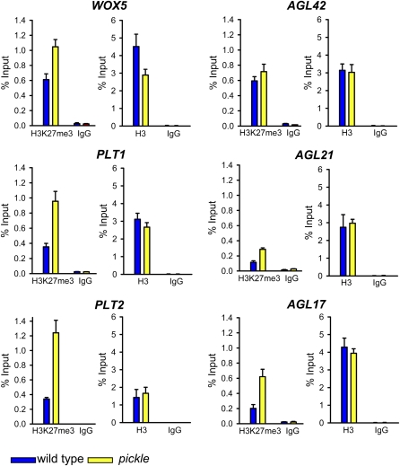
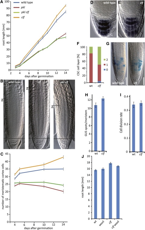
The chromatin modifying Polycomb group (PcG) and trithorax group (trxG) proteins are central regulators of cell identity that maintain a tightly controlled balance between cell proliferation and cell differentiation. The opposing activities of PcG and trxG proteins ensure the correct expression of specific transcriptional programs at defined developmental stages. Here, we report that the chromatin remodeling factor PICKLE (PKL) and the PcG protein CURLY LEAF (CLF) antagonistically determine root meristem activity. Whereas loss of PKL function caused a decrease in meristematic activity, loss of CLF function increased meristematic activity. Alterations of meristematic activity in pkl and clf mutants were not connected with changes in auxin concentration but correlated with decreased or increased expression of root stem cell and meristem marker genes, respectively. Root stem cell and meristem marker genes are modified by the PcG-mediated trimethylation of histone H3 on lysine 27 (H3K27me3). Decreased expression levels of root stem cell and meristem marker genes in pkl correlated with increased levels of H3K27me3, indicating that root meristem activity is largely controlled by the antagonistic activity of PcG proteins and PKL.


Figures

Figure 1.
Figure 1.
Genes Only Detected in Enriched Domains or Cell Types from the Root Are Downregulated in pkl and pkl pkr2 Roots. (A) Microarray analysis of seedling, inflorescence, rosette, and root tissues reveals root-specific expression of genes with decreased expression in roots of pkl pkr2 seedlings. Numbers of microarrays used for this analysis are indicated on right side of panel. Analysis of tissue specificity of differentially expressed genes was performed in Genevestigator (Zimmermann et al., 2004). Differentially expressed genes in pkl pkr2 were identified using previously published data sets and following previously published procedures (Aichinger et al., 2009). (B) Genes only detected in enriched domains or cell types from the root are downregulated in pkl and pkl pkr2 mutants. Differentially expressed genes in pkl and pkl pkr2 were identified using previously published data sets and following previously published procedures (Aichinger et al., 2009). SLR, signal log ratio. Error bars indicate se. [See online article for color version of this figure.]
Figure 2.
Figure 2.
Loss of PKL and PKR2 Causes Reduced Root Growth Independent of Embryonic Trait Expression. (A) Phenotype of wild-type, pkl, pkr2, and pkl pkr2 seedlings at 5 DAG. Seedlings were stained with the embryonic lipid staining dye Fat Red. Bar = 5 mm. (B) Root length analysis of wild-type, pkl, pkr2, and pkl pkr2 seedlings from 3 to 7 DAG. n ≥ 34 for each data point; error bars indicate se. (C) Root length analysis of wild-type pkl, pkr2, and pkl pkr2 seedlings at 5 DAG in presence or absence of GA. n ≥ 34 for each data point; error bars indicate se. (D) Expression analysis of embryonic regulators LEC1, FUS3, ABI3, and AGL15 in wild-type-like and embryonic roots of pkl and pkl pkr2 mutants at 5 DAG. wt, wild type. (E) Expression analysis of LEC1 in primary roots of amiRNA_LEC1 lines in pkl pkr2 background and pkl pkr2 at 5 DAG. (F) Root length analysis of five independent amiRNA_LEC1 lines in pkl pkr2 background at 7 DAG. n ≥ 20 for each data point; error bars indicate se.
Figure 3.
Figure 3.
pkl and pkl pkr2 Mutants Have Reduced Meristem Size. (A) Cell size of differentiated root epidermis cell is not changed in pkl mutants in comparison to the wild type (wt) but significantly reduced in pkl pkr2 (two-tailed Student’s t test, P = 0.034). Error bars indicate se. (B) Microscopy images of Arabidopsis RMs at 5 DAG. The QC is marked in yellow. The QC is surrounded by stem cells; stem cells giving rise to endodermis and cortex cell files are marked in red. Columella stem cells are marked in green. The cortical cell file is framed by white lines. The meristem is marked by lines, and the triangle marks the transition from the meristem to the elongation zone. Bar = 50 μm. (C) Number of meristematic cortex cells in wild-type, pkl, pkr2, and pkl pkr2 seedlings at various time points; meristem size of pkl and pkl pkr2 from 3 DAG onwards is significantly different from the wild type and the pkr2 single mutant (Student’s t test, P < 0.001); n ≥ 48 for each data point; error bars indicate se. (D) Expression of PKLpro:PKL-GFP in pkl pkr2 roots at 5 DAG. Arrowhead marks QC. Bar = 50 μm.
Figure 4.
Figure 4.
Cell Division Rate of Meristematic Cortex Cells in pkl Roots Is Unchanged. (A) GUS-staining cells in pkl and wild-type roots expressing the CYCLINB1;1pro:CDB-GUS marker at 5 DAG. Bar = 50 μm. (B) Quantification of GUS spots per meristem in pkl and wild-type (wt) roots. n = 50; error bars indicate se. (C) Quantification of cell division rate in pkl and wild-type roots as number of GUS spot per meristematic cortex cell. Error bars indicate se.
Figure 5.
Figure 5.
pkl Seedling Roots Have Reduced Root Stem Cell Activity. (A) to (C) Lugol stained roots of the wild type (A) and pkl ([B] and [C]) at 5 DAG. Asterisk marks QC cells. Error heads point at stained stem cells. Bar = 25 μm. (D) Quantification of stem cell layers in wild-type and pkl roots at 5 DAG. Col, n = 30; pkl, n = 89. (E) Quantitative RT-PCR analysis of AGL42, AGL21, AGL17, PLT1, PLT2, WOX5, and SHR in wild-type and pkl roots at 5 DAG. Wild-type-like roots correspond to pkl roots that do not display the embryonic root phenotype. Error bars indicate se. (F) Expression of DR5pro:GUS in wild-type and pkl roots. Bar = 50 μm. (G) Root length analysis of wild-type (wt), pkl, plt1 plt2, and plt1 plt2 pkl mutants at 5 DAG. Col, n = 143; pkl, n = 86; plt1 plt2, n = 139; pkl plt1 plt2, n = 107. Error bars indicate se.
Figure 6.
Figure 6.
CLF and PKL Antagonistically Regulate Root Stem Cell and Meristem Marker Genes. (A) Quantitative RT-PCR analysis of AGL42, AGL21, AGL17, PLT1, PLT2, WOX5, and SHR in wild-type, pkl, clf, and pkl clf roots at 5 DAG. Error bars indicate se. Asterisks indicate transcript levels that were significantly altered compared with the wild type (one-tailed Student’s test, P < 0.01). (B) Expression of WOX5pro:GFP in wild-type, pkl, clf, and pkl clf roots. Bar = 50 μm. (C) Expression of DR5pro:GUS in wild-type and clf roots. Bar = 50 μm.
Figure 7.
Figure 7.
Root Stem Cell and Meristem Marker Genes Are PcG Target Genes. ChIP analysis of H3K27me3 and H3 levels at AGL42, AGL21, AGL17, PLT1, PLT2, and WOX5 in wild-type and pkl roots at 5 DAG. Nonspecific IgG antibodies served as a negative control. Quantitative ChIP PCR was performed with four replicates, and quantification of the results is shown as the recovery of the percentage of the input. Error bars indicate se. [See online article for color version of this figure.]
Figure 8.
Figure 8.
CLF and PKL Antagonistically Regulate RM Activity. (A) Root length analysis of wild-type pkl, clf, and pkl clf seedlings from 2 to 14 DAG. n (2, 3, 8, 9, 10, and 12 DAG) ≥ 22 seedlings; n (5 and 14 DAG) ≥ 50 seedling. Error bars indicate se. (B) Microscopy images of wild-type, clf, and pkl clf RMs at 5 DAG. The meristem is marked by lines, and the triangle marks the transition from the meristem to the elongation zone. Bar = 50 μm. (C) Number of meristematic cortex cells in wild-type, pkl, clf, and pkl clf seedlings at various time points. n ≥ 48 for each data point; error bars indicate se. (D) and (E) Lugol-stained roots of the wild type (C) and clf (D) at 5 DAG. Asterisk marks QC cells. Bar = 50 μm. (F) Quantification of columella stem cell (CSC) layers in wild-type (wt) and clf roots at 5 DAG. Col, clf, n = 46. (G) GUS-staining cells in clf and wild-type roots expressing the CYCLINB1;1pro:CDB-GUS marker at 5 DAG. Bar = 50 μm. (H) Quantification of GUS spots per meristem in clf and wild-type roots. n = 50; error bars indicate se. (I) Quantification of cell division rate in clf and wild-type roots as number of GUS spot per meristematic cortex cell. Error bars indicate se. (J) Root length analysis of wox5, clf, and clf wox5 compared to the wild type at 5 DAG. Col, n = 80; wox5, n = 73, clf, n = 81; clf wox5, n = 91. Root lengths of clf and clf wox5 are significantly different from the wild type (P < 0.001). Root length of clf is significantly different from clf wox5 (P < 0.05). Student’s t test P < 0.05. Error bars indicate se.

References

    1. Aichinger E., Villar C.B., Farrona S., Reyes J.C., Hennig L., Köhler C. (2009). CHD3 proteins and polycomb group proteins antagonistically determine cell identity in Arabidopsis. PLoS Genet. 5: e1000605. - PMC - PubMed
    1. Aida M., Beis D., Heidstra R., Willemsen V., Blilou I., Galinha C., Nussaume L., Noh Y.S., Amasino R., Scheres B. (2004). The PLETHORA genes mediate patterning of the Arabidopsis root stem cell niche. Cell 119: 109–120 - PubMed
    1. Baker S.P., Grant P.A. (2007). The SAGA continues: Expanding the cellular role of a transcriptional co-activator complex. Oncogene 26: 5329–5340 - PMC - PubMed
    1. Blilou I., Xu J., Wildwater M., Willemsen V., Paponov I., Friml J., Heidstra R., Aida M., Palme K., Scheres B. (2005). The PIN auxin efflux facilitator network controls growth and patterning in Arabidopsis roots. Nature 433: 39–44 - PubMed
    1. Bouazoune K., Brehm A. (2006). ATP-dependent chromatin remodeling complexes in Drosophila. Chromosome Res. 14: 433–449 - PubMed

Publication types

MeSH terms