. 2011 Mar;23(3):1171-85.
doi: 10.1105/tpc.110.075093. Epub 2011 Mar 25.

The membrane mucin Msb2 regulates invasive growth and plant infection in Fusarium oxysporum

Affiliations

The membrane mucin Msb2 regulates invasive growth and plant infection in Fusarium oxysporum

Elena Pérez-Nadales et al. Plant Cell. 2011 Mar.

Abstract

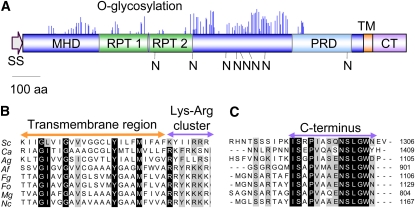
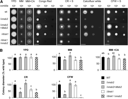
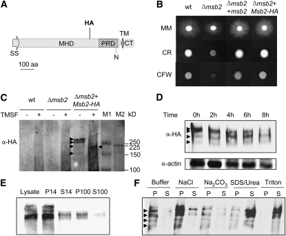
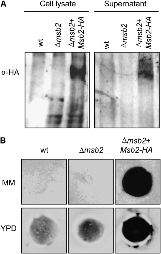
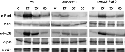
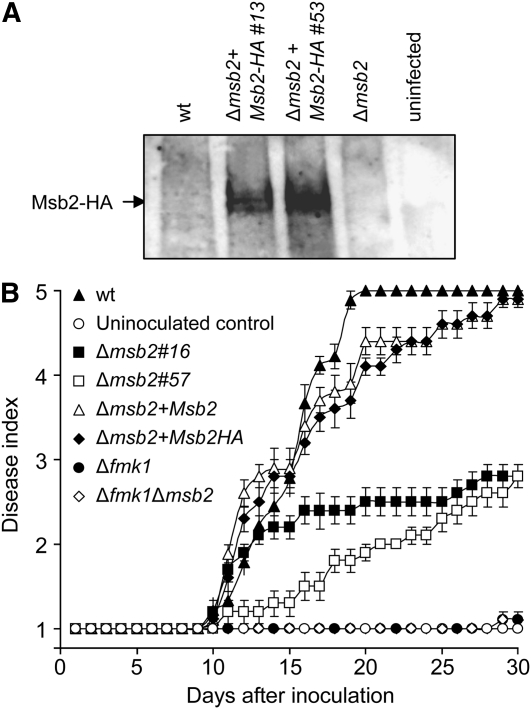
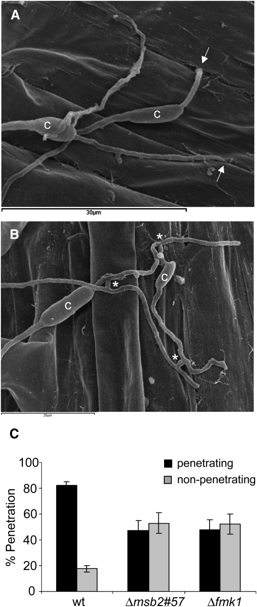
Fungal pathogenicity in plants requires a conserved mitogen-activated protein kinase (MAPK) cascade homologous to the yeast filamentous growth pathway. How this signaling cascade is activated during infection remains poorly understood. In the soil-borne vascular wilt fungus Fusarium oxysporum, the orthologous MAPK Fmk1 (Fusarium MAPK1) is essential for root penetration and pathogenicity in tomato (Solanum lycopersicum) plants. Here, we show that Msb2, a highly glycosylated transmembrane protein, is required for surface-induced phosphorylation of Fmk1 and contributes to a subset of Fmk1-regulated functions related to invasive growth and virulence. Mutants lacking Msb2 share characteristic phenotypes with the Δfmk1 mutant, including defects in cellophane invasion, penetration of the root surface, and induction of vascular wilt symptoms in tomato plants. In contrast with Δfmk1, Δmsb2 mutants were hypersensitive to cell wall targeting compounds, a phenotype that was exacerbated in a Δmsb2 Δfmk1 double mutant. These results suggest that the membrane mucin Msb2 promotes invasive growth and plant infection upstream of Fmk1 while contributing to cell integrity through a distinct pathway.


Figures

Figure 1.
Figure 1.
F. oxysporum Msb2 Is a Structural Ortholog of the S. cerevisiae Msb2 Mucin. (A) Schematic representation of the F. oxysporum Msb2 protein. Shown are the N-terminal signal sequence (SS), the extracellular Ser/Thr/Pro-rich MHD with imperfect repeats (RPT), the PRD, the TM, and the CT. O-glycosylation sites (blue peaks) and N-glycosylation sites (N) are represented as predicted by NetOGlyc 3.1 and NetNGlyc 1.0, respectively. Each peak represents the score value calculated for a Ser or Thr residue in the sequence above the limiting threshold. aa, amino acids. (B) and (C) Amino acid sequence alignment of the TM (B) and C-terminal intracellular regions (C) of the putative Msb2 orthologs of S. cerevisiae (Sc), C. albicans (Ca), A. gossypii (Ag), A. fumigatus (Af), F. graminearum (Fg), F. oxysporum (Fo), M. oryzae (Mg), and N. crassa (Nc). Highly conserved residues are shaded in black; moderately conserved residues are shaded in gray.
Figure 2.
Figure 2.
Msb2 Contributes to Hyphal Growth under Conditions of Nutrient Limitation and Cell Integrity Stress. (A) Colony phenotype of the indicated strains grown on yeast peptone Glc (YPD), minimal medium (MM), MM supplemented with 1% (w/v) casaminoacids (MM+CA), YPD supplemented with 50 μg/mL Congo Red (CR), or 40 μg/mL Calcofluor White (CFW) in the absence or presence of 1 M sorbitol (S). Plates were spot-inoculated with the indicated amount of microconidia, incubated for five (YPD, MM, and MM+CA) or three (CR and CFW) days at 28°C and scanned. Bar = 1 cm. wt, wild type. (B) Colony diameter on the indicated media was measured after 5 d and plotted relative to the wild-type (WT) strain (100%). Error bars represent standard deviations of colony diameters calculated from five plates. Values with the same letter are not significantly different according to the Mann-Whitney test (P ≤ 0.05).
Figure 3.
Figure 3.
Msb2 Is a Glycosylated Membrane Protein. (A) Schematic representation of the Msb2-HA protein carrying the HA epitope at amino acid (aa) residue 722 located in the MHD region. Symbols and abbreviations are as in Figure 1A. (B) Colony phenotype of the indicated strains grown on YPD medium in the absence or presence of CR or CFW. Experimental conditions are as in Figure 2. wt, wild type. (C) Deglycosylation of Msb2. Cell lysates from the indicated strains germinated for 15 h in potato dextrose broth (PDB) and subsequently transferred for 8 h onto MM plates were incubated in the absence or presence of TMSF, separated by SDS-PAGE, and subjected to immunoblot analysis with monoclonal α-HA antibody. Hybridizing bands are marked by arrowheads. Molecular masses of prestained markers from Bio-Rad and Amersham (M1 and M2, respectively) are indicated to the right. (D) Immunoblot analysis of cell lysates from the Δmsb2+Msb2-HA strain germinated as described in (C) and transferred onto MM plates for the indicated time periods. (E) Subcellular localization of Msb2-HA. Immunoblot analysis of cell lysates of the Δmsb2+Msb2-HA strain obtained as described in (C) and separated by centrifugation. Crude lysate, supernatant (S), and pellet (P) fractions are shown. 14, 14,000g; 100, 100,000g. (F) P14 fraction analysis. Treatments were as follows: lysis buffer alone (Buffer) or with 0.5 M NaCl, 100 mM Na2CO3 at pH 11, 5% SDS + 8 M urea, or 1% Triton.
Figure 4.
Figure 4.
Msb2 Is Shed from the Cell Surface. (A) Immunoblot with α-HA antibody of cell lysates and culture supernatants of the indicated strains. wt, wild type. (B) Colony inmunoblot assay. Microconidia of the indicated strains were germinated for 15 h in PDB, harvested and washed twice in water, transferred onto 0.2-μm pore size filters placed over a plate of MM or YPD overlaid with a nitrocellulose filter, and incubated for 8 h at 28°C. The 0.2-μm filters with the colonies were removed, and nitrocellulose membranes were washed with running water and subjected to immunoblot with α-HA antibody.
Figure 5.
Figure 5.
Msb2 Contributes to Phosphorylation of the Fmk1 and Hog1 MAPKs. Transfer to solid medium induces a transient Msb2-dependent increase in phosphorylation of Fmk1 and Hog1. Total protein extracts from the indicated strains germinated for 15 h in PDB and transferred onto MM plates for the indicated time periods (min) were subjected to immunoblot analysis with anti-phospho-p44/42 MAPK antibody (α-P-erk) that only detects the phosphorylated form of Fmk1and anti-p44/p42 MAPK antibody (α-ERK) or with anti-phospho-p38 MAPK antibody (α-P-p38) that only detects the phosphorylated form of Hog1 and anti-p38 MAPK antibody (α-p38). A monoclonal α-actin antibody was used as loading control. wt, wild type.
Figure 6.
Figure 6.
Msb2 Modulates the Expression of Fmk1-Regulated Effector Genes. mRNA abundance of the indicated genes was measured 6 h after transfer of the strains to MM plates using real-time qPCR. Relative expression levels represent mean cycle threshold values normalized to actin gene expression levels and relative to expression values in the wild-type (WT) strain. Bars represent standard errors calculated from three biological replicates. Values with the same letter are not significantly different according to the Mann-Whitney test (P ≤ 0.05).
Figure 7.
Figure 7.
Msb2 Is Expressed during the Early Stages of Infection and Contributes to Virulence on Tomato Plants. (A) Total protein extracts were obtained from tomato roots 48 h after inoculation with microconidia of the indicated strains or from uninoculated roots and submitted to immunoblot analysis with α-HA antibody. wt, wild type. (B) Incidence of Fusarium wilt on tomato plants inoculated with the indicated strains. Severity of disease symptoms was recorded using an index ranging from 1 (healthy plant) to 5 (dead plant). Error bars represent standard errors calculated from 20 plants.
Figure 8.
Figure 8.
Msb2 Contributes to Penetration of Tomato Roots. (A) and (B) Scanning electron microscopy analysis of tomato roots, 24 h after inoculation with microconidia of the F. oxysporum f. sp lycopersici wild-type (A) or Δmsb2 (B) strain. (A) shows fungal hyphae entering the root through openings at intercellular junctions. Arrows point to penetration events. (B) shows multiple hyphal fusion events, indicated by asterisks. c, conidium. (C) Tomato roots inoculated with the indicated strains and incubated for 24 h were analyzed by scanning electron microscopy. Bars indicate the percentage of fungal germlings showing at least one hypha penetrating the root. In each experiment, 20 germinated conidia per strain were surveyed. Error bars represent standard errors calculated from four independent experiments. Values with the same letter are not significantly different according to the Mann-Whitney test (P ≤ 0.05). wt, wild type.

References

    1. Almeida I.C., Ferguson M.A., Schenkman S., Travassos L.R. (1994). Lytic anti-alpha-galactosyl antibodies from patients with chronic Chagas’ disease recognize novel O-linked oligosaccharides on mucin-like glycosyl-phosphatidylinositol-anchored glycoproteins of Trypanosoma cruzi. Biochem. J. 304: 793–802 - PMC - PubMed
    1. Armstrong G.M., Armstrong J.K. (1981). Formae speciales and races of Fusarium oxysporum causing wilt diseases. Fusarium: Diseases, Biology and Taxonomy, Cook R., (University Park, PA: Penn State University Press; ), pp. 391–399
    1. Bendtsen J.D., Nielsen H., von Heijne G., Brunak S. (2004). Improved prediction of signal peptides: SignalP 3.0. J. Mol. Biol. 340: 783–795 - PubMed
    1. Bermejo C., Rodríguez E., García R., Rodríguez-Peña J.M., Rodríguez de la Concepción M.L., Rivas C., Arias P., Nombela C., Posas F., Arroyo J. (2008). The sequential activation of the yeast HOG and SLT2 pathways is required for cell survival to cell wall stress. Mol. Biol. Cell 19: 1113–1124 - PMC - PubMed
    1. Bishop C.D., Cooper R.M. (1983). An ultrastructural study of root invasion of three vascular wilt diseases. Physiol. Mol. Plant Pathol. 22: 15–27

Publication types

MeSH terms

Substances

LinkOut - more resources