. 2011 May 1;186(9):5367-75.
doi: 10.4049/jimmunol.1003438. Epub 2011 Mar 25.

Chronic exposure to a TLR ligand injures hematopoietic stem cells

Affiliations

Chronic exposure to a TLR ligand injures hematopoietic stem cells

Brandt L Esplin et al. J Immunol. .

Abstract

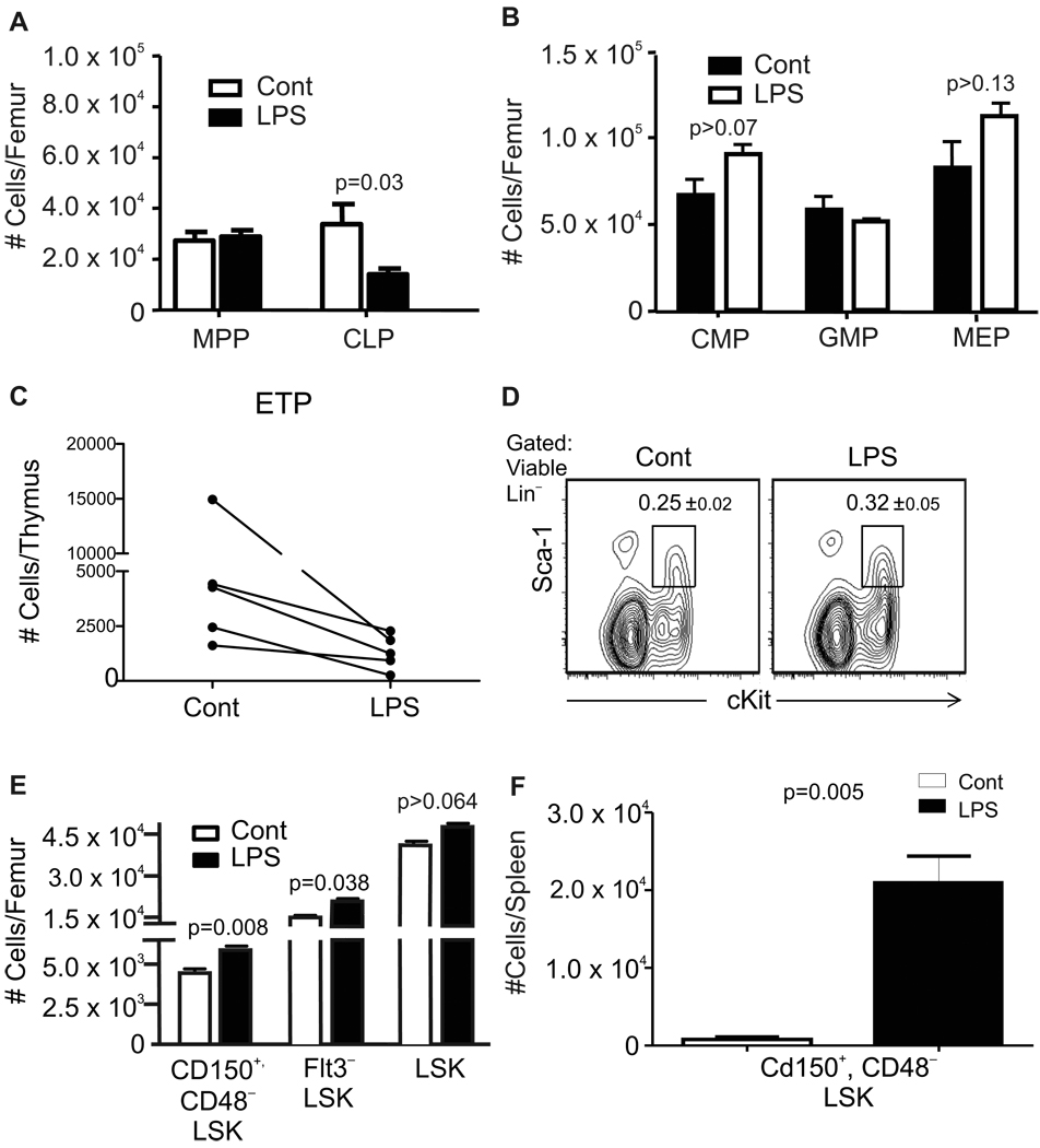
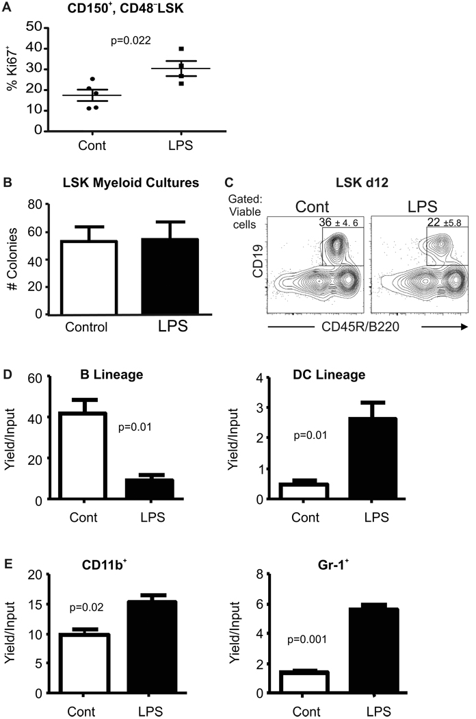
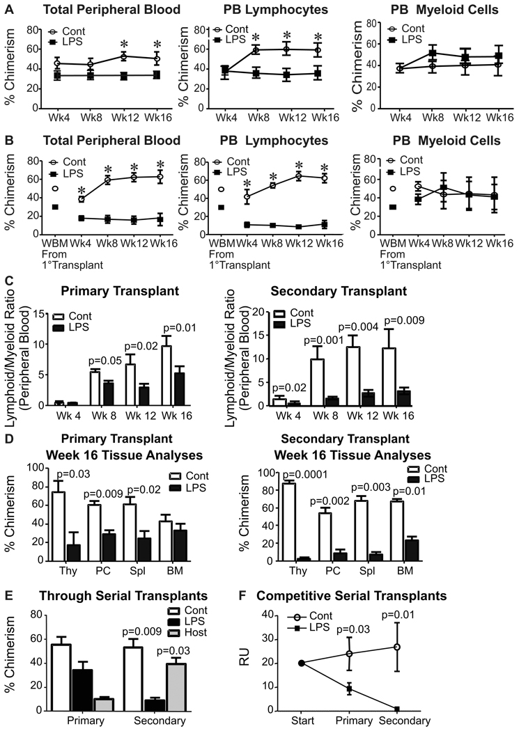
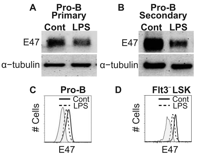
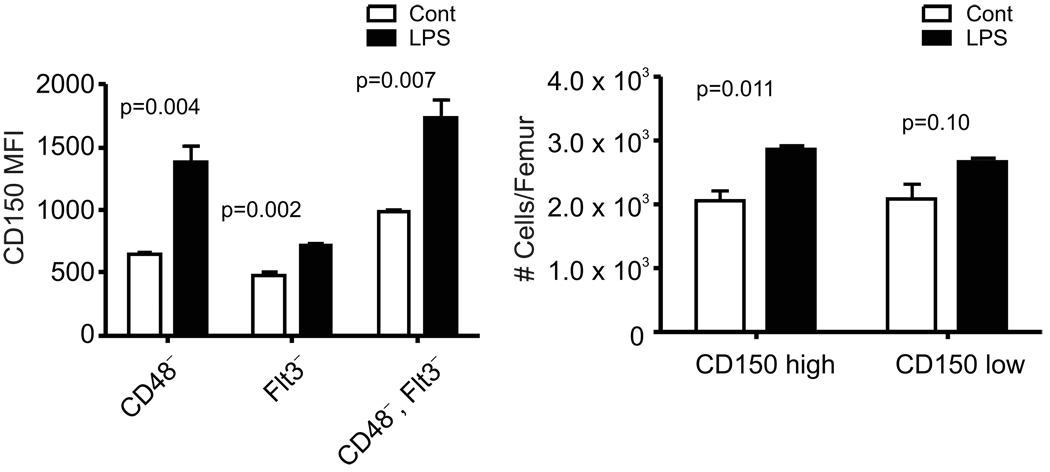
Hematopoietic stem cells (HSC) can be harmed by disease, chemotherapy, radiation, and normal aging. We show in this study that damage also occurs in mice repeatedly treated with very low doses of LPS. Overall health of the animals was good, and there were relatively minor changes in marrow hematopoietic progenitors. However, HSC were unable to maintain quiescence, and transplantation revealed them to be myeloid skewed. Moreover, HSC from treated mice were not sustained in serial transplants and produced lymphoid progenitors with low levels of the E47 transcription factor. This phenomenon was previously seen in normal aging. Screening identified mAbs that resolve HSC subsets, and relative proportions of these HSC changed with age and/or chronic LPS treatment. For example, minor CD150(Hi)CD48(-) populations lacking CD86 or CD18 expanded. Simultaneous loss of CD150(Lo/-)CD48(-) HSC and gain of the normally rare subsets, in parallel with diminished transplantation potential, would be consistent with age- or TLR-related injury. In contrast, HSC in old mice differed from those in LPS-treated animals with respect to VCAM-1 or CD41 expression and lacked proliferation abnormalities. HSC can be exposed to endogenous and pathogen-derived TLR ligands during persistent low-grade infections. This stimulation might contribute in part to HSC senescence and ultimately compromise immunity.


Conflict of interest statement

Disclosures

The authors have no financial conflicts of interest.

Figures

Figure 1
Figure 1. Slight perturbation of hematopoiesis during TLR ligand exposure
Mice were given daily 6µg injections of LPS, or PBS for 4–6 weeks before harvest of bone marrow and flow cytometry analyses (Gating illustrated in Supplemental Fig. 1B). A, Multipotent progenitors (MPP) defined as LinSca-1+cKitHiFlt3+ and LinSca-1+cKitLoIL-7Rα+ common lymphoid progenitors (CLP) were evaluated in the same samples. B, LinSca-1cKitHiCD34LoFcγRII/IIILo common myeloid progenitors (CMP), LinSca-1cKitHiFcγRII/III+CD34+ granulocyte-monocyte progenitor (GMP) and Lin Sca-1 cKitHi FcγRII/III CD34 megakaryocyte-erythroid progenitors (MEP) were enumerated in separate experiments. Additional values are given in Table 1, and it is noteworthy that numbers of total nucleated cells in the marrow were unaffected. These data are representative of four independent experiments with 4–6 mice per group, and are given as mean values ± SEM. C, Early thymocyte progenitors gated as Lin c-KitHi CD44Hi CD25 cells were enumerated and the results from five independent experiments are shown as individual plots. D, The interferon regulated Sca-1 antigen was not up-regulated on cells in the primitive LSK fraction of bone marrow. E, Absolute numbers of cells in the HSC enriched Flt3LSK fraction and the Lin Sca-1+ KitHiCD48 CD150+ stem cell subset were significantly increased, although this was not the case for the larger LSK category. F, Absolute numbers of Lin Sca-1+ KitHiCD48CD150+ stem cells in the spleen are shown for one of three independent experiments that gave very similar results (p=<0.005).
Figure 2
Figure 2. Primitive hematopoietic cells are altered by chronic LPS exposure
A, Cell cycling activity in CD150+ CD48 LSK in control and LPS treated marrow was assessed by flow cytometry for the Ki-67 proliferation antigen. B, LSK were sorted from mice at the end of LPS or PBS exposure and placed in myeloid supporting Methocel cultures. Average numbers of colonies were counted 10 days later. C, The same fractions were placed in defined, lymphoid supporting liquid cultures for 12 days before flow cytometry. Boxes show percentages of B220+CD19+ lymphocytes that were generated; (p=0.02). This data is representative of that found in four independent experiments with 5–8 mice per group. All error values depict SEM. D, Common lymphoid progenitors (CLP) defined as in Supplemental Fig. 1B were recovered from the two groups of mice after 4–6 weeks of chronic exposure and sorted to high purity. These were placed in defined lymphoid supporting cultures (see Experimental Procedures) for 8 days before harvest and flow cytometry. Yields of CD19+B220+ B-lineage lymphocytes and CD11c+ dendritic cells ±SEM per input progenitor were calculated. E, Common myeloid progenitors (CMP) sorted as shown in Supplemental Fig. 1B were placed in myeloid supporting cultures and their potential for generation of myeloid marker bearing cells was determined 8 days later by flow cytometry. Absolute yields of myeloid cells ±SEM per input progenitor are shown. Similar results were obtained in four independent experiments with at least four culture wells per group. Statistical significance was determined by t-test analysis.
Figure 3
Figure 3. Low-grade inflammation causes age-related changes in long-term HSC
Competitive serial marrow transplants were constructed as described in the Experimental Procedures. A, Peripheral blood was analyzed at 4 week intervals to determine hematopoietic contributions of PBS and LPS treated marrow after primary transplantation. Total peripheral blood chimerism is shown in the left panel, while CD11bGr-1CD3+/CD19+ lymphocytes and CD3CD19CD11b+ myeloid chimerism is indicated in the middle and right panels, respectively. B, Secondary transplants were initiated with chimeric marrow from primary transplants, as indicated. C, The same data was used to calculate lymphoid to myeloid cell ratios of donor type cells in peripheral blood from primary, as well as secondary transplants. D, Tissues recovered from these secondary transplant recipients at 16 weeks were analyzed for percentages of chimerism by flow cytometry. E, Marrow recovered was studied by flow cytometry to determine what proportions of cells in the primitive LSK fraction were derived from the original LPS treated animals, the PBS treated control mice or radio-resistant host mice. F, Repopulating units (RU) of control (CD45.1+) and LPS-treated (CD45.2+) marrow were calculated through serial transplantations initiated with equal numbers of competing whole marrow cells to determine the ability of both donor types to repetitively maintain their RU as an indication of HSC self-renewal capacity (41). This data represents pooled averages of total bone marrow chimerism after 16-week serial transplantation interval analyses from four independent experiments. Each one was conducted with not less than six recipient animals per group. Error bars depict SEM and asterisks reflect statistically significant differences.
Figure 4
Figure 4. Chronic TLR stimulation leads to decreased E2A protein abundance in pro-B and primitive cells
Western blot analysis comparing E47 protein levels in pro-B cells generated from control or LPS treated marrow after primary (A) and secondary (B) transplants. α-tubulin served as an internal protein control for this assay. The data is representative of three independent experiments per time point. Two independent flow cytometry analyses from separate laboratories assessed intracellular E47 protein levels in individual pro-B (C) and HSC-enriched Flt3LSK cells (D) after secondary transplantation. Plots are representative of 3 independent experiments with 5–6 mice per analysis. Statistical significance was determined by t-test analysis.
Figure 5
Figure 5. Repeated exposure to LPS increases densities of CD150 and numbers of CD150Hi HSC in bone marrow
Marrow was harvested from mice given daily injections of saline or LPS for four-six weeks. Subsets of HSC in the LSK gate illustrated in Supplemental Fig. 1B were resolved on the basis of CD48 and/or Flt3 expression. A, Densities of CD150 are reflected in mean fluorescence intensities (MFI). B, HSC identified as CD150+ CD48 LSK were subdivided into CD150Lo and CD150Hi fractions (See Supplemental Fig. 1B) that were enumerated in control and LPS treated mice.
Figure 6
Figure 6. Monoclonal antibody staining resolves HSC subsets altered by LPS treatment and/or aging
The indicated antibodies were used to characterize CD48 LSK in marrow of mice repeatedly injected with LPS or allowed to age naturally. Note that both of these conditions resulted in a shift to high CD150 expression on HSC (see also Fig. 5). These results are representative of more than three independent experiments with each reagent. Additionally, CD41 staining was confirmed with two different fluorochromes (PE or FITC). Mean percentages of labeled cells ± S.E. from one of three representative experiments are given within the boxes.

References

    1. Binder D, Fehr J, Hengartner H, Zinkernagel RM. Virus-induced transient bone marrow aplasia: major role of interferon-alpha/beta during acute infection with the noncytopathic lymphocytic choriomeningitis virus. J. Exp. Med. 1997;185:517–530. - PMC - PubMed
    1. Kurtzman GJ, Platanias L, Lustig L, Frickhofen N, Young NS. Feline parvovirus propagates in cat bone marrow cultures and inhibits hematopoietic colony formation in vitro. Blood. 1989;74:71–81. - PubMed
    1. Nakao S, Lai CJ, Young NS. Dengue virus, a flavivirus, propagates in human bone marrow progenitors and hematopoietic cell lines. Blood. 1989;74:1235–1240. - PubMed
    1. Zeldis JB, Mugishima H, Steinberg HN, Nir E, Gale RP. In vitro hepatitis B virus infection of human bone marrow cells. J. Clin. Invest. 1986;78:411–417. - PMC - PubMed
    1. Alexaki A, Wigdahl B. HIV-1 infection of bone marrow hematopoietic progenitor cells and their role in trafficking and viral dissemination. PLoS. Pathog. 2008;4:e1000215. - PMC - PubMed

Publication types

LinkOut - more resources