. 2011 Jun;193(11):2875-9.
doi: 10.1128/JB.00134-11. Epub 2011 Mar 25.

Role of the Nfo and ExoA apurinic/apyrimidinic endonucleases in radiation resistance and radiation-induced mutagenesis of Bacillus subtilis spores

Affiliations

Role of the Nfo and ExoA apurinic/apyrimidinic endonucleases in radiation resistance and radiation-induced mutagenesis of Bacillus subtilis spores

Ralf Moeller et al. J Bacteriol. 2011 Jun.

Abstract

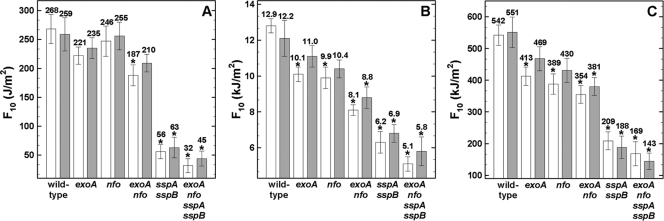
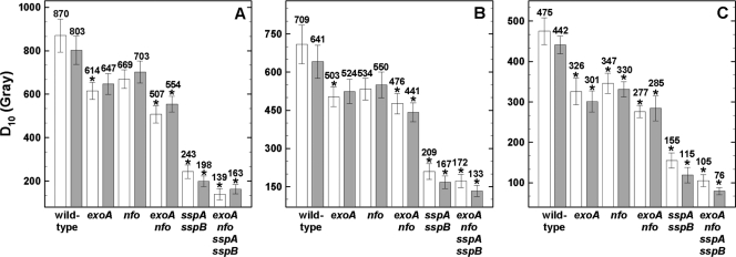
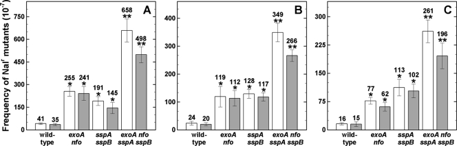
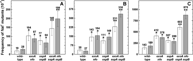
The roles of DNA repair by apurinic/apyrimidinic (AP) endonucleases alone, and together with DNA protection by α/β-type small acid-soluble spore proteins (SASP), in Bacillus subtilis spore resistance to different types of radiation have been studied. Spores lacking both AP endonucleases (Nfo and ExoA) and major SASP were significantly more sensitive to 254-nm UV-C, environmental UV (>280 nm), X-ray exposure, and high-energy charged (HZE)-particle bombardment and had elevated mutation frequencies compared to those of wild-type spores and spores lacking only one or both AP endonucleases or major SASP. These findings further implicate AP endonucleases and α/β-type SASP in repair and protection, respectively, of spore DNA against effects of UV and ionizing radiation.


Figures

Fig. 1.
Fig. 1.
Spore resistance to standard germicidal 254-nm UV-C (A), environmentally relevant 280- to 400-nm UV-(A+B) (B), and 320- to 400-nm UV-A (C) radiation. Spores were irradiated as air-dried monolayers (white bars) or in water (gray bars), and F10 values are expressed as averages ± standard deviations (n = 3) as described in the text. Asterisks indicate F10 values that were significantly different (P ≤ 0.05) from those for wild-type spores. Note the differences in the y axes between panels A, B, and C.
Fig. 2.
Fig. 2.
Spore resistance to X rays (A) and high-energy charged helium (B) and iron (C) ions. Spores were irradiated as air-dried monolayers (white bars) or in water (gray bars), and D10 values are expressed as averages ± standard deviations (n = 3) as described in the text. Asterisks indicate D10 values that were significantly different (P ≤ 0.05) compared to wild-type spores. Note the differences in the y axes between panels A, B, and C.
Fig. 3.
Fig. 3.
Mutation frequencies to Nalr of dormant spores of different strains irradiated with 100 J/m2 of 254-nm UV-C (A), 10 kJ/m2 of UV-(A+B) (B), and 250 kJ/m2 of UV-A (C) radiation. Spores were exposed as air-dried monolayers (white bars) or in water (gray bars), and values are averages ± standard deviations for triplicate determinations in three separate experiments as described in the text. One asterisk indicates a mutation frequency that was significantly different from that of wild-type spores, and two asterisks indicate a mutation frequency that was also significantly different from that of αβ spores (Student's t test; P ≤ 0.05). Note the differences in the y axes in the different panels. The spontaneous mutation frequency to Nalr of the tested strains ranged from 1.6 × 10⁻⁷ to 8.3 × 10⁻⁷, in good agreement with previous data (10).
Fig. 4.
Fig. 4.
Mutation frequencies to Nalr of dormant spores of different strains irradiated with 250 Gy of X rays (A) and high-energy helium ions and (B) and iron ions (C). Spores were exposed as air-dried monolayers (white bars) or in water (gray bars), and values are averages ± standard deviations for triplicate determinations in three separate experiments as described in the text. One asterisk indicates a mutation frequency that was significantly different from that of wild-type spores, and two asterisks indicate a mutation frequency that was also significantly different from that of αβ spores (Student's t test; P ≤ 0.05). Note the differences in the y axes in the different panels. The spontaneous mutation frequency to Nalr of the tested strains ranged from 1.6 × 10⁻⁷ to 8.3 × 10⁻⁷, in good agreement with previous work (10).

References

    1. Barraza-Salas M., et al. 2010. Effects of forespore-specific overexpression of apurinic/apyrimidinic endonuclease Nfo on the DNA-damage resistance properties of Bacillus subtilis spores. FEMS Microbiol. Lett. 302:159–165 - PubMed
    1. Blaisdell J. O., Harrison L., Wallace S. S. 2001. Base excision repair processing of radiation-induced clustered DNA lesions. Radiat. Prot. Dosimetry 97:25–31 - PubMed
    1. Boiteux S., Guillet M. 2004. Abasic sites in DNA: repair and biological consequences in Saccharomyces cerevisiae. DNA Repair 3:1–12 - PubMed
    1. Breimer L. H. 1988. Ionizing radiation-induced mutagenesis. Br. J. Cancer 57:6–18 - PMC - PubMed
    1. Cadet J., Douki T., Ravanat J. L. 2010. Oxidatively generated base damage to cellular DNA. Free Radic. Biol. Med. 49:9–21 - PubMed

Publication types

LinkOut - more resources