. 2011 May;193(10):2408-17.
doi: 10.1128/JB.01262-10. Epub 2011 Mar 25.

Unexpected diversity of chlorite dismutases: a catalytically efficient dimeric enzyme from Nitrobacter winogradskyi

Affiliations

Unexpected diversity of chlorite dismutases: a catalytically efficient dimeric enzyme from Nitrobacter winogradskyi

Georg Mlynek et al. J Bacteriol. 2011 May.

Abstract

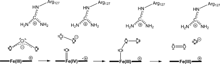
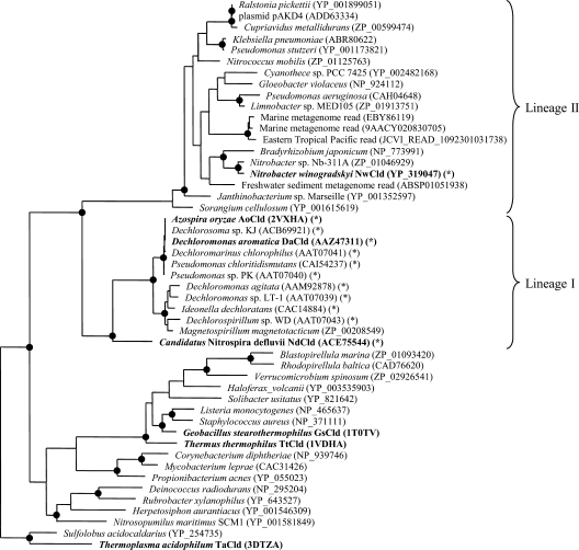
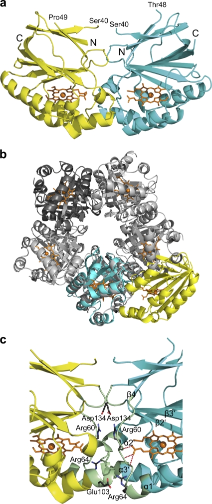
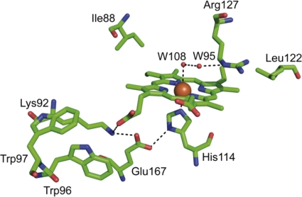
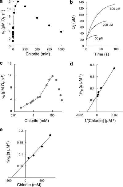
Chlorite dismutase (Cld) is a unique heme enzyme catalyzing the conversion of ClO(2)(-) to Cl(-) and O(2). Cld is usually found in perchlorate- or chlorate-reducing bacteria but was also recently identified in a nitrite-oxidizing bacterium of the genus Nitrospira. Here we characterized a novel Cld-like protein from the chemolithoautotrophic nitrite oxidizer Nitrobacter winogradskyi which is significantly smaller than all previously known chlorite dismutases. Its three-dimensional (3D) crystal structure revealed a dimer of two identical subunits, which sharply contrasts with the penta- or hexameric structures of other chlorite dismutases. Despite a truncated N-terminal domain in each subunit, this novel enzyme turned out to be a highly efficient chlorite dismutase (K(m) = 90 μM; k(cat) = 190 s(-1); k(cat)/K(m) = 2.1 × 10(6) M(-1) s(-1)), demonstrating a greater structural and phylogenetic diversity of these enzymes than was previously known. Based on comparative analyses of Cld sequences and 3D structures, signature amino acid residues that can be employed to assess whether uncharacterized Cld-like proteins may have a high chlorite-dismutating activity were identified. Interestingly, proteins that contain all these signatures and are phylogenetically closely related to the novel-type Cld of N. winogradskyi exist in a large number of other microbes, including other nitrite oxidizers.


Figures

Fig. 1.
Fig. 1.
Proposed reaction mechanism of chlorite dismutase starting with the attack of anionic chlorite at ferric heme b. After formation of the Fe(III)-chlorite complex (not shown), heterolytic cleavage of the O-Cl bond leads to the formation of hypochlorite and the redox intermediate compound I [oxoiron(IV) porphyrin cation radical]. Finally, upon nucleophilic attack of anionic hypochlorite at the ferryl oxygen, compound I is reduced to the resting state and dioxygen and chloride are released. In addition, the scheme shows the putative role of conserved Arg127 in the orientation and stabilization of the substrate and the postulated intermediate(s).
Fig. 2.
Fig. 2.
Maximum-likelihood phylogenetic tree based on the amino acid sequences of selected Clds and Cld-like proteins. Names printed in boldface represent proteins with known crystal structures. Validated and catalytically efficient Clds are marked with an asterisk. The proposed phylogenetic lineages I (canonical Clds, mainly from PCRB) and II (NwCld and related proteins) are delimited by curly brackets. Black circles on tree nodes symbolize high-parsimony bootstrap support (≥90%) based on 100 iterations. Accession numbers are indicated for all sequences.
Fig. 3.
Fig. 3.
NwCld subunit and structural comparison with canonical NdCld. (a) Ribbon representation of one NwCld subunit. The N-terminal domain is shown in red, whereas the C-terminal domain is shown in cyan. The N and C termini are labeled, and the heme b is presented as orange sticks, with iron shown as an orange sphere. The labeling of secondary structure elements follows the labeling used for NdCld as shown in panel b. (b) Ribbon representation of one subunit of NdCld from “Ca. Nitrospira defluvii.” Secondary structure elements are labeled according to the method of Kostan et al. (22). Heme group and iron are presented as in panel a. (c) A subunit of NdCld (shown in green and gray) superimposed on a subunit of NwCld (shown in red and cyan). The orientation of the subunits is the same as in panels a and b. Structural elements missing in the subunit structure of NwCld compared to the NdCld subunit structure are depicted in gray.
Fig. 4.
Fig. 4.
NwCld dimer structure and interface. (a) Ribbon representation of the NwCld dimer viewed perpendicular to the vertical 2-fold symmetry axis. Subunits are shown in different colors. The heme group is shown as an orange stick model in either subunit. The iron is displayed as an orange sphere. The N and C termini are labeled. Parts of the structure between Ser40 and Thr48 in one subunit and Ser40 and Pro49 in the other subunit could not be modeled in the structure due to weak electron density in these regions. (b) Superposition of an NwCld dimer with an NdCld pentamer (shown in gray), with one subunit of NwCld (cyan) spatially aligned to one NdCld monomer. Note the different location of the second NwCld subunit (yellow) compared to those of the subunits of NdCld, illustrating differences in the interfaces between the subunits of dimeric and pentameric Clds. To more easily follow the packing of subunits in the NdCld pentamer, one of its subunits is depicted in dark gray. (c) Detailed view of the NwCld dimer interface. Residues involved in interactions between the two subunits of the NwCld holoenzyme are shown in pale green. Side chains of amino acids involved in the formation of salt bridges are shown as sticks, with carbon, oxygen, and nitrogen atoms depicted in gray, red, and blue, respectively. Hemes are presented as orange stick models, with heme irons shown as orange spheres. Interactions of heme propionate with the protein backbone are visualized by red dashes.
Fig. 5.
Fig. 5.
Active site of NwCld. The illustration shows selected residues involved in heme b binding and in the catalytic mechanism of chlorite dismutation. Carbon, oxygen, and nitrogen atoms are depicted in green, red, and blue, respectively. The heme iron and water molecules (W) are shown as orange spheres. The hydrogen bonding network spanning from Arg127 to Glu167 is visualized by dashed lines.
Fig. 6.
Fig. 6.
Kinetics of chlorite dismutation activity by NwCld. (a) Plot of the initial rate (v0) of molecular oxygen evolution as a function of chlorite concentration. Points represent averages of three measurements. Applied conditions were as follows: 50 mM phosphate buffer (pH 7.0), 20 nM NwCld, 30°C. (b) Selected time traces at different chlorite concentrations. Conditions were as for panel a. (c) Semilogarithmic plot of the data in panel a. Double-rectangular hyperbolar fits are shown in gray. (d) Double reciprocal plot of v0 versus chlorite concentration for the determination of kinetic parameters. (e) Dixon plot for the determination of Ki.

References

    1. Adams P. D., et al. 2010. PHENIX: a comprehensive Python-based system for macromolecular structure solution. Acta Crystallogr. D Biol. Crystallogr. 66:213–221 - PMC - PubMed
    1. Balk M., van Gelder T., Weelink S. A., Stams A. J. M. 2008. (Per)chlorate reduction by the thermophilic bacterium, Moorella perchloratireducens sp. nov., isolated from an underground gas storage. Appl. Environ. Microbiol. 74:403–409 - PMC - PubMed
    1. Bender K. S., Rice M. R., Fugate W. H., Coates J. D., Achenbach L. A. 2004. Metabolic primers for detection of (per)chlorate-reducing bacteria in the environment and phylogenetic analysis of cld gene sequences. Appl. Environ. Microbiol. 70:5651–5658 - PMC - PubMed
    1. Bendtsen J. D., Nielsen H., von Heijne G., Brunak S. 2004. Improved prediction of signal peptides: SignalP 3.0. J. Mol. Biol. 340:783–795 - PubMed
    1. Bendtsen J. D., Nielsen H., Widdick D., Palmer T., Brunak S. 2005. Prediction of twin-arginine signal peptides. BMC Bioinform. 6:167 - PMC - PubMed

Publication types

Associated data

LinkOut - more resources