

Muscle-specific expression of insulin-like growth factor 1 improves outcome in Lama2Dy-w mice, a model for congenital muscular dystrophy type 1A
- PMID: 21441569
- PMCID: PMC3098729
- DOI: 10.1093/hmg/ddr126
Muscle-specific expression of insulin-like growth factor 1 improves outcome in Lama2Dy-w mice, a model for congenital muscular dystrophy type 1A
Abstract
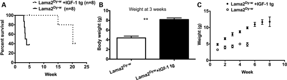
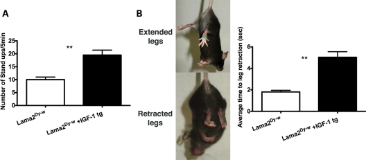
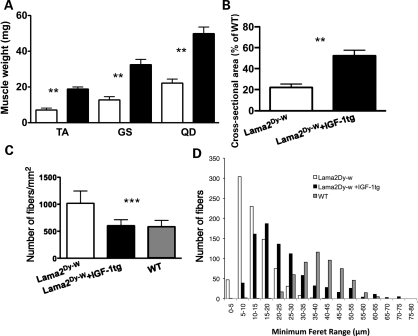
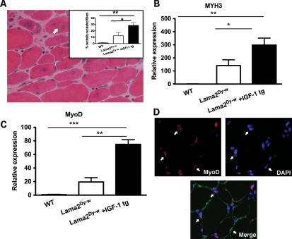
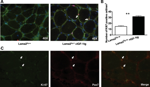
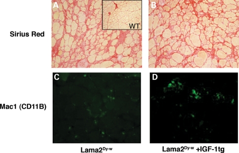
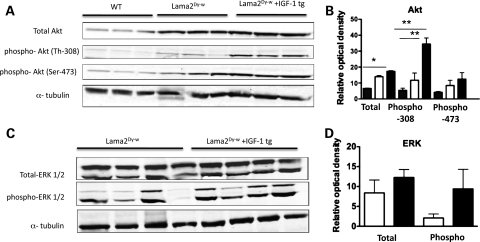
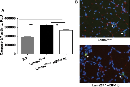
MDC1A, the second most prevalent form of congenital muscular dystrophy, results from laminin-α2 chain deficiency. This disease is characterized by extensive muscle wasting that results in extremely weak skeletal muscles. A large percentage of children with MDC1A are faced with respiratory as well as ambulatory difficulties. We investigated the effects of overexpressing insulin-like growth factor-1 (IGF-1) as a potential therapeutic target for the disease in the Lama2(Dy-w) mouse, a model that closely resembles human MDC1A. IGF-1 transgenic Lama2(Dy-w) mice showed increased survivability, body weight and muscle weight. In addition, these mice showed better ability to stand up on their hind limbs: a typical exploratory behavior seen in healthy mice. Histology and immunohistochemistry analyses revealed increased regenerative capacity and proliferation in IGF-1 transgenic Lama2(Dy-w) muscles. Western blot analysis showed increased phosphorylation of Akt and ERK1/2, both known to enhance myogenesis. Additionally, we saw increases in the expression of the regeneration markers MyoD, myogenin and embryonic myosin (myosin heavy chain 3, MYH3). We conclude that overexpression of IGF-1 in Lama2(Dy-w) mice increases lifespan and improves their overall wellbeing mainly through the restoration of impaired muscle regeneration, as fibrosis or inflammation was not impacted by IGF-1 in this disease model. Our results demonstrate that IGF-1 has a promising therapeutic potential in the treatment of MDC1A.
Figures
Similar articles
-
Triggering regeneration and tackling apoptosis: a combinatorial approach to treating congenital muscular dystrophy type 1 A.Hum Mol Genet. 2013 Nov 1;22(21):4306-17. doi: 10.1093/hmg/ddt280. Epub 2013 Jun 16. Hum Mol Genet. 2013. PMID: 23773998
-
Laminin-111 protein therapy reduces muscle pathology and improves viability of a mouse model of merosin-deficient congenital muscular dystrophy.Am J Pathol. 2012 Apr;180(4):1593-602. doi: 10.1016/j.ajpath.2011.12.019. Epub 2012 Feb 6. Am J Pathol. 2012. PMID: 22322301 Free PMC article.
-
Laminin-111 improves muscle repair in a mouse model of merosin-deficient congenital muscular dystrophy.Hum Mol Genet. 2014 Jan 15;23(2):383-96. doi: 10.1093/hmg/ddt428. Epub 2013 Sep 5. Hum Mol Genet. 2014. PMID: 24009313 Free PMC article.
-
Laminin-α2 Chain-Deficient Congenital Muscular Dystrophy: Pathophysiology and Development of Treatment.Curr Top Membr. 2015;76:31-60. doi: 10.1016/bs.ctm.2015.05.002. Curr Top Membr. 2015. PMID: 26610911 Review.
-
Improving Reproducibility of Phenotypic Assessments in the DyW Mouse Model of Laminin-α2 Related Congenital Muscular Dystrophy.J Neuromuscul Dis. 2017;4(2):115-126. doi: 10.3233/JND-170217. J Neuromuscul Dis. 2017. PMID: 28550268 Free PMC article. Review.
Cited by
-
Transcriptomic Profile of Primary Culture of Skeletal Muscle Cells Isolated from Semitendinosus Muscle of Beef and Dairy Bulls.Int J Mol Sci. 2020 Jul 7;21(13):4794. doi: 10.3390/ijms21134794. Int J Mol Sci. 2020. PMID: 32645861 Free PMC article.
-
The dystrophin-glycoprotein complex in the prevention of muscle damage.J Biomed Biotechnol. 2011;2011:210797. doi: 10.1155/2011/210797. Epub 2011 Oct 5. J Biomed Biotechnol. 2011. PMID: 22007139 Free PMC article. Review.
-
Current understanding and treatment of cardiac and skeletal muscle pathology in laminin-α2 chain-deficient congenital muscular dystrophy.Appl Clin Genet. 2019 Jul 3;12:113-130. doi: 10.2147/TACG.S187481. eCollection 2019. Appl Clin Genet. 2019. PMID: 31308722 Free PMC article.
-
The non-muscle ADF/cofilin-1 controls sarcomeric actin filament integrity and force production in striated muscle laminopathies.Cell Rep. 2021 Aug 24;36(8):109601. doi: 10.1016/j.celrep.2021.109601. Cell Rep. 2021. PMID: 34433058 Free PMC article.
-
Quantitative proteomic analysis reveals metabolic alterations, calcium dysregulation, and increased expression of extracellular matrix proteins in laminin α2 chain-deficient muscle.Mol Cell Proteomics. 2014 Nov;13(11):3001-13. doi: 10.1074/mcp.M113.032276. Epub 2014 Jul 3. Mol Cell Proteomics. 2014. PMID: 24994560 Free PMC article.
References
-
- Muntoni F., Voit T. The congenital muscular dystrophies in 2004: a century of exciting progress. Neuromuscul. Disord. 2004;14:635–649. - PubMed
-
- Muntoni F., Bertini E., Bonnemann C., Brockington M., Brown S., Bushby K., Fiszman M., Korner C., Mercuri E., Merlini L., et al. 98th ENMC International Workshop on Congenital Muscular Dystrophy (CMD), 7th Workshop of the International Consortium on CMD, 2nd Workshop of the MYO CLUSTER project GENRE. 26–28th October, 2001, Naarden, The Netherlands. Neuromuscul. Disord. 2002;12:889–896. - PubMed
-
- Colognato H., Yurchenco P.D. Form and function: the laminin family of heterotrimers. Dev. Dyn. 2000;218:213–234. - PubMed
-
- Langenbach K.J., Rando T.A. Inhibition of dystroglycan binding to laminin disrupts the PI3K/AKT pathway and survival signaling in muscle cells. Muscle Nerve. 2002;26:644–653. - PubMed
-
- Laprise P., Poirier E.M., Vezina A., Rivard N., Vachon P.H. Merosin-integrin promotion of skeletal myofiber cell survival: differentiation state-distinct involvement of p60Fyn tyrosine kinase and p38alpha stress-activated MAP kinase. J. Cell Physiol. 2002;191:69–81. - PubMed
Publication types
MeSH terms
Substances
Supplementary concepts
Grants and funding
LinkOut - more resources
Full Text Sources
Other Literature Sources
Medical
Molecular Biology Databases
Miscellaneous
