Review
. 2011 Jul 1;3(7):a003707.
doi: 10.1101/cshperspect.a003707.

Spliceosome structure and function

Affiliations
Review

Spliceosome structure and function

Cindy L Will et al. Cold Spring Harb Perspect Biol. .

Abstract

Pre-mRNA splicing is catalyzed by the spliceosome, a multimegadalton ribonucleoprotein (RNP) complex comprised of five snRNPs and numerous proteins. Intricate RNA-RNA and RNP networks, which serve to align the reactive groups of the pre-mRNA for catalysis, are formed and repeatedly rearranged during spliceosome assembly and catalysis. Both the conformation and composition of the spliceosome are highly dynamic, affording the splicing machinery its accuracy and flexibility, and these remarkable dynamics are largely conserved between yeast and metazoans. Because of its dynamic and complex nature, obtaining structural information about the spliceosome represents a major challenge. Electron microscopy has revealed the general morphology of several spliceosomal complexes and their snRNP subunits, and also the spatial arrangement of some of their components. X-ray and NMR studies have provided high resolution structure information about spliceosomal proteins alone or complexed with one or more binding partners. The extensive interplay of RNA and proteins in aligning the pre-mRNA's reactive groups, and the presence of both RNA and protein at the core of the splicing machinery, suggest that the spliceosome is an RNP enzyme. However, elucidation of the precise nature of the spliceosome's active site, awaits the generation of a high-resolution structure of its RNP core.


Figures
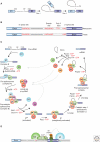
Figure 1.
Figure 1.
Pre-mRNA splicing by the U2-type spliceosome. (A) Schematic representation of the two-step mechanism of pre-mRNA splicing. Boxes and solid lines represent the exons (E1, E2) and the intron, respectively. The branch site adenosine is indicated by the letter A and the phosphate groups (p) at the 5′ and 3′ splice sites, which are conserved in the splicing products, are also shown. (See facing page for legend.) (B) Conserved sequences found at the 5′ and 3′ splice sites and branch site of U2-type pre-mRNA introns in metazoans and budding yeast (S. cerevisiae). Y = pyrimidine and R = purine. The polypyrimidine tract is indicated by (Yn). (C) Canonical cross-intron assembly and disassembly pathway of the U2-dependent spliceosome. For simplicity, the ordered interactions of the snRNPs (indicated by circles), but not those of non-snRNP proteins, are shown. The various spliceosomal complexes are named according to the metazoan nomenclature. Exon and intron sequences are indicated by boxes and lines, respectively. The stages at which the evolutionarily conserved DExH/D-box RNA ATPases/helicases Prp5, Sub2/UAP56, Prp28, Brr2, Prp2, Prp16, Prp22 and Prp43, or the GTPase Snu114, act to facilitate conformational changes are indicated. (D) Model of interactions occurring during exon definition.
Figure 2.
Figure 2.
Protein composition and snRNA secondary structures of the major human spliceosomal snRNPs. All seven Sm proteins (B/B’, D3, D2, D1, E, F, and G) or LSm proteins (Lsm2-8) are indicated by “Sm” or “LSm” at the top of the boxes showing the proteins associated with each snRNP. The U4/U6.U5 tri-snRNP contains two sets of Sm proteins and one set of LSm proteins.
Figure 3.
Figure 3.
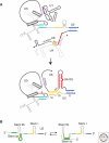
Dynamic network of RNA-RNA interactions in the spliceosome. (A) Exon sequences are indicated by grey boxes and intron sequences by a thin black line. snRNAs are shown schematically (secondary structure as observed in mammals) in grey or black, with those regions engaging in base pairing interactions (indicated by short lines) highlighted in color (not drawn to scale). The 5′ end of the snRNAs is indicated by a black dot. Solely loop 1 of the U5 snRNA is shown. During the transition from a precatalytic spliceosome (upper diagram) to a catalytically activated spliceosome (lower diagram) U1 and U4 are displaced, and U6 and U2 engage in novel base pairing interactions. (B) Conformational toggling of the yeast U2 snRNA. Two mutually exclusive stem structures (stem IIa and stem IIc) are thought to form within the U2 snRNA at different stages of splicing. Solely the 5′ end of the U2 snRNA is shown schematically.
Figure 4.
Figure 4.
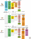
Compositional dynamics of the yeast spliceosome. Proteins identified by mass spectrometry in S. cerevisiae B, Bact, and C spliceosomal complexes are shown. Proteins are grouped according to their function or association with an snRNP, protein complex or spliceosomal complex. The relative abundance of the indicated proteins is indicated by light (substoichiometric) or dark (stoichiometric) lettering. (Reprinetd, with permission, from Fabrizio et al. 2009 [© Elsevier].)
Figure 5.
Figure 5.
Three dimensional EM structure of the U4/U6.U5 tri-snRNP and localization of functionally important tri-snRNP proteins. (A) 3D reconstructions of the human U5 and U4/U6 snRNPs and tri-snRNP, and fitting of U5 and U4/U6 into the tri-snRNP 3D map (adapted, with permission, from Sander et al. 2006 [© Elsevier]). The head domain of U5 (highlighted blue) appears to be flexible and it is positioned in the U5 snRNP 3D reconstruction shown in a manner favorable for fitting into the tri-snRNP 3D map. (B) Left, representative 2D class average of the affinity purified S. cerevisiae U4/U6.U5 tri-snRNP as visualized by negative-stain electron microscopy after mild fixation using the Grafix protocol. The main structural domains are indicated. Right, cartoon model of the yeast tri-snRNP. Area corresponding to the U5 and U4/U6 snRNPs and the linker region are shaded grey, orange, or yellow, respectively. The position of the carboxyl terminus of several tri-snRNP proteins is indicated (adapted from Häcker et al. 2008).
Figure 6.
Figure 6.
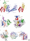
Structures of the U4 snRNA 5′ stem-loop complexed with Prp31 and the 15.5K protein, the RNAse H-like domain of Prp8, and the U1 snRNP. (A) Left, ribbon diagram of hPrp31 (residues 78-333)(purple) and the 15.5K protein (red) complexed with the human U4 snRNA 5'stem loop (nts 20-52) (gold and green). RNA elements absent in the 15.5K-U4 RNA binary complex (because of the shorter RNA used) (shown at right) are highlighted in green. A disordered loop of hPrp31 in the ternary complex and an unstructured region of the U4 pentaloop in the binary complex are indicated by a dashed lines. Mutations at positions A194 and A216 (shown as cyan-colored space-filling models) are linked to retinitis pigmentosa. (See facing page for legend). hPrp31 binds to one region of the composite binding platform formed by 15.5K and the U4 snRNA by a lock-and-key type mechanism, and another region of the RNA via an induced fit mechanism. (Reprinted, with permission, from Liu et al. 2007 [© AAAS].) (B) Ribbon diagram (left) and space filling model (right) of the S. cerevisiae Prp8 protein (residues 1827-2092). Left, the mixed β-sheet and two α-helices typical of RNAse H domains are highlighted, red and purple, respectively. The Prp8-specific β-hairpin and α-helices are colored magenta and green, respectively. Residues comprising the active site in RNAse H (corresponding to Asp1853 and Asp1854 in yeast Prp8) are indicated by sticks and the 310 helix that is crosslinked to the 5′ splice site is highlighted cyan blue. Right, modeling of the pre-mRNA (exon and intron nucleotides, brown and beige, respectively and 5′ss phosphate, black) into the Prp8 RNAse-like domain space filling model. Site of Prp8 crosslinks to the 5′ss is encompassed by a dashed line and the predicted active site, gold. The Brr 2 interacting region is shown in green. The sites of Prp8 mutations (amino acid residue indicated) suppressing 5′ss (blue), 3′ss (green), polypyrimidine tract (purple) and U4 cs1 (magenta) mutations are indicated. (C) Left, ribbon diagram of the U1 snRNP containing the Sm proteins and the 70K and C proteins. The U1 snRNA, with stem-loop (SL) 1, 3 and 4, and the 5′ end indicated, is shaded grey. Orange spheres indicate anomalous peaks from SeMet (introduced at the indicated amino acid position) in U1-70K. Middle, ribbon diagram of the Sm proteins (E, F, G, D1, D2, D3, B) and seven nucleotide Sm site RNA, with the experimental electron density map (contoured at 1σ). Right, Ribbon diagram with experimental electron density map (contoured at 1σ) of the interaction of the 5′ end of U1 snRNA with a neighboring complex (orange) which mimics the 5′ss of the pre-mRNA. (Reprinted, with permission, from Newman and Nagai 2010 [© Elsevier]; originally Pomeranz-Krummel et al. 2009 [© Macmillan].)
Figure 7.
Figure 7.
Structural dynamics of the yeast spliceosome as visualized by EM and localization of the pre-mRNA in the human B complex. (A) Class average of electron microscopy images of negatively stained, affinity-purified human B complexes (right). Sketch of the B complex showing regions where the 5′ exon, 3′ exon, intron and SF3b155 protein were mapped by immuno-EM, and the likely location of components of the A complex and tri-snRNP. (Adapted from Wolf et al. 2009 [© Nature Publishing Group].) (B) Electron microscopy of negatively-stained, affinity-purified S. cerevisiae B, Bact, and C complexes. Two prominent class averages of each complex are shown, with the maximum dimension indicated later. (Adapted, with permission, from Fabrizio et al. 2009 [© Elsevier].)

References

    1. Abelson J 2008. Is the spliceosome a ribonucleoprotein enzyme? Nat Struct Mol Biol 15: 1235–1237 - PubMed
    1. Alcid EA, Jurica MS 2008. A protein-based EM label for RNA identifies the location of exons in spliceosomes. Nat Struct Mol Biol 15: 213–215 - PMC - PubMed
    1. Aronova A, Bacíková D, Crotti LB, Horowitz DS, Schwer B 2007. Functional interactions between Prp8, Prp18, Slu7, and U5 snRNA during the second step of pre-mRNA splicing. RNA 13: 1437–1444 - PMC - PubMed
    1. Ast G 2004. How did alternative splicing evolve? Nat Rev 5: 773–782 - PubMed
    1. Azubel M, Wolf SG, Sperling J, Sperling R 2004. Three-dimensional structure of the native spliceosome by cryo-electron microscopy. Mol Cell 15: 833–839 - PubMed

Publication types

MeSH terms

LinkOut - more resources