

Cell-extracellular matrix interactions in normal and diseased skin
- PMID: 21441589
- PMCID: PMC3062212
- DOI: 10.1101/cshperspect.a005124
Cell-extracellular matrix interactions in normal and diseased skin
Abstract
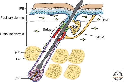
Mammalian skin comprises a multi-layered epithelium, the epidermis, and an underlying connective tissue, the dermis. The epidermal extracellular matrix is a basement membrane, whereas the dermal ECM comprises fibrillar collagens and associated proteins. There is considerable heterogeneity in ECM composition within both epidermis and dermis. The functional significance of this extends beyond cell adhesion to a range of cell autonomous and nonautonomous processes, including control of epidermal stem cell fate. In skin, cell-ECM interactions influence normal homeostasis, aging, wound healing, and disease. Disturbed integrin and ECM signaling contributes to both tumor formation and fibrosis. Strategies for manipulating cell-ECM interactions to repair skin defects and intervene in a variety of skin diseases hold promise for the future.
Figures

References
-
- Aden N, Nuttall A, Shiwen X, de Winter P, Leask A, Black CM, Denton CP, Abraham DJ, Stratton RJ 2010. Epithelial cells promote fibroblast activation via IL-1α in systemic sclerosis. J Invest Dermatol PMID: 20445556 - PubMed
-
- Asano Y, Ihn H, Jinnin M, Mimura Y, Tamaki K 2006a. Involvement of αvβ5 integrin in the establishment of autocrine TGF-β signaling in dermal fibroblasts derived from localized scleroderma. J Invest Dermatol 126: 1761–1769 - PubMed
-
- Benitah SA, Frye M, Glogauer M, Watt FM 2005. Stem cell depletion through epidermal deletion of Rac1. Science 309: 933–935 - PubMed
Publication types
MeSH terms
Grants and funding
LinkOut - more resources
Full Text Sources
Other Literature Sources
Medical