Review
. 2011 May 1;3(5):a004960.
doi: 10.1101/cshperspect.a004960.

Tenascins and the importance of adhesion modulation

Affiliations
Review

Tenascins and the importance of adhesion modulation

Ruth Chiquet-Ehrismann et al. Cold Spring Harb Perspect Biol. .

Abstract

Tenascins are a family of extracellular matrix proteins that evolved in early chordates. There are four family members: tenascin-X, tenascin-R, tenascin-W, and tenascin-C. Tenascin-X associates with type I collagen, and its absence can cause Ehlers-Danlos Syndrome. In contrast, tenascin-R is concentrated in perineuronal nets. The expression of tenascin-C and tenascin-W is developmentally regulated, and both are expressed during disease (e.g., both are associated with cancer stroma and tumor blood vessels). In addition, tenascin-C is highly induced by infections and inflammation. Accordingly, the tenascin-C knockout mouse has a reduced inflammatory response. All tenascins have the potential to modify cell adhesion either directly or through interaction with fibronectin, and cell-tenascin interactions typically lead to increased cell motility. In the case of tenascin-C, there is a correlation between elevated expression and increased metastasis in several types of tumors.


Figures

Figure 1.
Figure 1.
Tenascins and fibronectin are only found in Chordates. The tenascin gene from amphioxus (Cephalochordata) encodes a predicted protein with the characteristic organization of tenascins: one or more EGF-like repeats near the amino terminus (yellow pentagons), FN3 domains (circles), and a carboxy-terminal fibrinogen-related domain (purple rectangle). FN3 domains with an RGD or KGD motif appropriately exposed for integrin binding are orange and those without are green. No fibronectin gene has been identified in amphioxus. In contrast, tunicates (Urochordata) have both a tenascin and a fibronectin-like gene. In mammals (adapted from human sequences here), there are four tenascin genes and a single fibronectin gene. In contrast, bony fish (Actinopterygii) have two fibronectin genes and the tenascin-C gene has duplicated. Representative proteins are illustrated here. Some are only predicted. In addition, some tenascins are known to have multiple splice variants that can result in different numbers of FN3 domains (see Fig. 2).
Figure 2.
Figure 2.
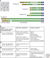
Main features of the four vertebrate tenascins. (A) Top left shows a tenascin-C hexabrachion revealed in an electron micrograph after rotary shadowing. Domain models of each tenascin family member are depicted for human and mouse orthologs as indicated by h (human) or m (mouse) within their fibrinogen-related domains (FReD). Heptad repeats for oligomerization are present close to the amino terminus indicated by a short black line in front of the EGF-like repeats shown in yellow and FN3 domains in dark green for the constant repeats, light green for FN3 repeats prone to alternative splicing, and orange for RGD-containing FN3 repeats. (B) Accession numbers on which the models in (A) are based and the chromosomal locations of the genes are given in the first column. Note that these protein sequences do not include certain extra repeats existing in rare splice variants. Main sites of expression are summarized as well as the major human or mouse disease associations found in the literature and cited in the main text.
Figure 3.
Figure 3.
Adhesion modulation by tenascin-C. Cells adopt different morphologies depending on the substratum to which they are adhering. Although the MCF7 epithelial cancer cells form cell layers with close cell–cell contacts typical of epithelia on fibronectin, they disperse and lose their cell-cell contacts on a tenascin-C substratum, a process termed epithelial-mesenchymal transition (or EMT) that is important in cancer invasion. In addition, the myogenic/osteogenic mouse cell line C2C12 reacts differently to these two substrata. As revealed by phalloidin staining the cells elaborate stress fibers on fibronectin while they concentrate F-actin in cell protrusions when plated on tenascin-C.
Figure 4.
Figure 4.
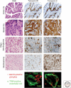
Tenascin-C and tenascin-W in cancer stroma. Immunostaining for tenascin-W and tenascin-C of sections of colon carcinoma (colon Ca) and breast carcinoma (breast Ca) reveal stromal staining in both cancer types (brown staining corresponding to the areas poor in nuclei visible in the corresponding H&E stained adjacent sections). In brain cancer (oligodendroglioma and glioblastoma) tenascin-W is found around blood vessels while tenascin-C is in addition also detected throughout the entire glioblastoma tissue and is secreted by these highly invasive cancer cells. The lowest panels reveal that tenascin-W and tenascin-C are expressed by different blood vessel cells: tenascin-W seems to be made by endothelial cells, whereas tenascin-C is made by the surrounding desmin-positive pericytes.

References

    1. Ambort D, Brellier F, Becker-Pauly C, Stocker W, Andrejevic-Blant S, Chiquet M, Sterchi EE 2010. Specific processing of tenascin-C by the metalloprotease meprinβ neutralizes its inhibition of cell spreading. Matrix Biol 29: 31–42 - PubMed
    1. Andjus PR, Bajic A, Zhu L, Schachner M, Strata P 2005. Short-term facilitation and depression in the cerebellum: Some observations on wild-type and mutant rodents deficient in the extracellular matrix molecule tenascin C. Ann N Y Acad Sci 1048: 185–197 - PubMed
    1. Apostolova I, Irintchev A, Schachner M 2006. Tenascin-R restricts posttraumatic remodeling of motoneuron innervation and functional recovery after spinal cord injury in adult mice. J Neurosci 26: 7849–7859 - PMC - PubMed
    1. Beiter K, Hiendlmeyer E, Brabletz T, Hlubek F, Haynl A, Knol C, Kirchner T, Jung A 2005. β-Catenin regulates the expression of tenascin-C in human colorectal tumors. Oncogene 24: 8200–8204 - PubMed
    1. Borsi L, Allemanni G, Gaggero B, Zardi L 1996. Extracellular pH controls pre-mRNA alternative splicing of tenascin-C in normal, but not in malignantly transformed, cells. Int J Cancer 66: 632–635 - PubMed

Publication types

LinkOut - more resources