. 2011 May 18;30(10):2057-70.
doi: 10.1038/emboj.2011.86. Epub 2011 Mar 25.

The cellular prion protein mediates neurotoxic signalling of β-sheet-rich conformers independent of prion replication

Affiliations

The cellular prion protein mediates neurotoxic signalling of β-sheet-rich conformers independent of prion replication

Ulrike K Resenberger et al. EMBO J. .

Abstract

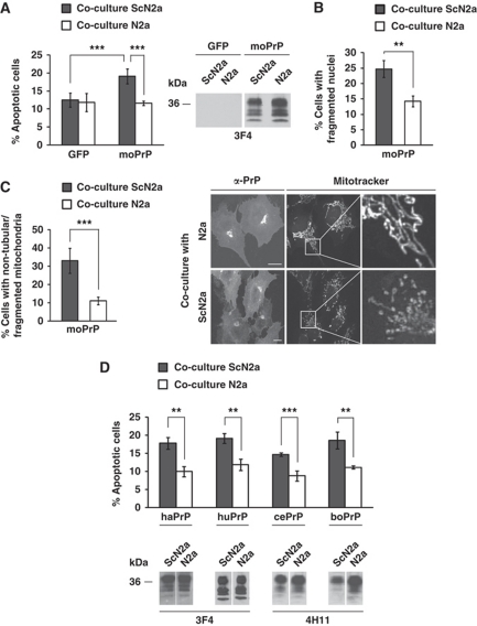
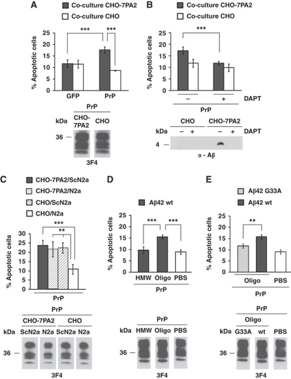
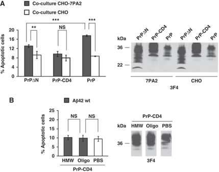
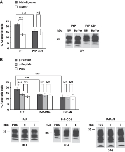
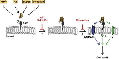
Formation of aberrant protein conformers is a common pathological denominator of different neurodegenerative disorders, such as Alzheimer's disease or prion diseases. Moreover, increasing evidence indicates that soluble oligomers are associated with early pathological alterations and that oligomeric assemblies of different disease-associated proteins may share common structural features. Previous studies revealed that toxic effects of the scrapie prion protein (PrP(Sc)), a β-sheet-rich isoform of the cellular PrP (PrP(C)), are dependent on neuronal expression of PrP(C). In this study, we demonstrate that PrP(C) has a more general effect in mediating neurotoxic signalling by sensitizing cells to toxic effects of various β-sheet-rich (β) conformers of completely different origins, formed by (i) heterologous PrP, (ii) amyloid β-peptide, (iii) yeast prion proteins or (iv) designed β-peptides. Toxic signalling via PrP(C) requires the intrinsically disordered N-terminal domain (N-PrP) and the GPI anchor of PrP. We found that the N-terminal domain is important for mediating the interaction of PrP(C) with β-conformers. Interestingly, a secreted version of N-PrP associated with β-conformers and antagonized their toxic signalling via PrP(C). Moreover, PrP(C)-mediated toxic signalling could be blocked by an NMDA receptor antagonist or an oligomer-specific antibody. Our study indicates that PrP(C) can mediate toxic signalling of various β-sheet-rich conformers independent of infectious prion propagation, suggesting a pathophysiological role of the prion protein beyond of prion diseases.


Conflict of interest statement

The authors declare that they have no conflict of interest.

Figures

Figure 1
Figure 1
Scrapie prions induce apoptosis and interfere with mitochondrial integrity in cells expressing homologous and heterologous PrPC. (A, B) SH-SY5Y cells expressing mouse (mo) PrP or GFP were co-cultured with ScN2a or N2a cells for 16 h. For quantification of apoptotic cell death, SH-SY5Y cells were fixed, permeabilized and stained for active caspase-3 (A) or fragmented nuclei (B). Expression of PrP was analysed by western blotting using the anti-PrP antibody 3F4 (middle panel). (C) Scrapie prions interfere with mitochondrial integrity. SH-SY5Y cells expressing moPrP were co-cultured with ScN2a or N2a cells for 16 h. Cells were stained with MitoTracker Red CMXRos to visualize mitochondria and analysed by fluorescence microscopy (right panel). Cells displaying an intact network of tubular mitochondria were classified as tubular. When this network was disrupted and mitochondria appeared predominantly spherical or rod-like, they were classified as fragmented. For quantification (left panel), the mitochondrial morphology of at least 500 cells per experiment was determined in a blinded manner. Quantifications were based on triplicates of at least three independent experiments. (D) PrPC mediates toxic signalling of heterologous PrPSc. SH-SY5Y cells transiently expressing hamster (ha), human (hu), cervid (ce) or bovine (bo) PrPC were co-cultured with ScN2a or N2a cells for 16 h. Shown is the percentage of apoptotic cells among transfected cells. Expression of transfected constructs was analysed by western blotting using the anti-PrP antibody 4H11 or 3F4. **P<0.005; ***P<0.0005.
Figure 2
Figure 2
Soluble oligomers of Aβ induce apoptosis in PrPC-expressing cells. (A, B) SH-SY5Y cells expressing the constructs indicated were co-cultivated with CHO-7PA2 or CHO cells for 16 h and apoptotic cell death in SH-SY5Y cells was determined as described in Figure 1A. (A) Soluble oligomers of Aβ secreted by stably transfected cells induce apoptosis in PrPC-expressing cells. GPI-anchored GFP was used as control. Expression of PrPC in CHO-7PA2 or CHO cells was analysed by western blotting using the 3F4 antibody (lower panel). (B) Inhibition of γ-secretase interferes with toxic effects of CHO-7PA2 cells. SH-SY5Y cells expressing PrPC were co-cultured with DAPT-treated (1 μM) CHO-7PA2 or CHO cells. Aβ present in the conditioned medium of CHO-7PA2 or CHO cells was analysed by immunoprecipitation followed by western blotting (lower panel). (C) SH-SY5Y cells expressing moPrPC were co-cultivated with the cell lines indicated. After16 h of co-cultivation, apoptotic cell death in SH-SY5Y cells was determined as described in Figure 1A. Expression of PrPC in co-cultured SH-SY5Y cells was analysed by western blotting using the 3F4 antibody (lower panel). (D) Synthetic Aβ42 oligomers are toxic to cells expressing PrPC. SH-SY5Y cells transiently expressing moPrPC were incubated in the presence of either oligomers (oligo) or high-molecular weight aggregates (HMW) of Aβ42 (500 nM each) for 12 h and apoptotic cell death was determined. Expression of PrPC was analysed by western blotting using the 3F4 antibody (lower panel). (E) Decreased toxicity of oligomers formed by mutant Aβ42 G33A. SH-SY5Y cells expressing moPrPC were incubated in the presence of Aβ42 G33A oligomers (500 nM) as described in (C). Expression of PrPC was monitored by western blotting using the anti-PrP antibody 3F4 (lower panel). **P<0.005; ***P<0.0005.
Figure 3
Figure 3
Toxic signalling of PrPC is dependent on the intrinsically disordered N-terminal domain and the C-terminal GPI anchor. (A) Apoptotic activity of soluble Aβ oligomers secreted by stably transfected CHO-7PA2 cells is compromised in cells expressing PrP mutants. SH-SY5Y cells expressing the PrP constructs indicated were co-cultivated with CHO-7PA2 or CHO cells and apoptotic cell death in SH-SY5Y cells was determined as described in Figure 1A. Expression of PrP constructs was controlled by western blotting using the 3F4 antibody (right panel). (B) Synthetic Aβ42 oligomers are not toxic to cells expressing PrP-CD4. SH-SY5Y cells expressing PrP-CD4 were incubated with either oligomers (oligo) or high-molecular weight aggregates (HMW) of Aβ42 (500 nM each) for 12 h and apoptotic cell death was determined. **P<0.005; ***P<0.0005; NS, non-significant. Expression of PrP-CD4 was analysed by western blotting using the 3F4 antibody (right panel).
Figure 4
Figure 4
PrPC mediates toxic signalling of an oligomeric yeast prion protein and of β-sheet-rich oligomers formed by a designed peptide. (A, B) β-sheet-rich conformers of completely different origin induce apoptosis via PrPC. SH-SY5Y cells expressing the PrP constructs indicated were incubated with either an oligomeric yeast prion protein (NM, 500 nM) for 7 h (A) or a synthetic β-peptide or α-peptide (200 nM each) for 12 h (B). Apoptotic cell death was determined as described in Figure 1A. Expression of the PrP constructs was controlled by western blotting using the 3F4 antibody. **P<0.005; ***P<0.0005; NS, non-significant.
Figure 5
Figure 5
Toxic signalling can be inhibited by a secreted version of the intrinsically disordered N-terminal domain of PrP. (A) The N-terminal domain of PrP mediates association with β-peptides. SH-SY5Y cells expressing the indicated PrP constructs were grown on cover slips and incubated for 2 h at 37°C with the β-peptide (200 nM). SH-SY5Y cells were fixed and stained with the polyclonal anti-PrP antibody A7 and the monoclonal anti-myc antibody 4A6, recognizing the β-peptide. Cell nuclei were visualized by DAPI. Scale bar, 10 μm. (B) Schematic presentation of PrP and the PrP/Fc fusion constructs analysed. PrPC/Fc, C-terminal domain of PrP fused to the Fc portion of human IgG1; PrPN/Fc, N-terminal domain of PrP fused to the Fc portion; SS/Fc, the ER signal sequence of PrP fused to the Fc portion; ER-SS, ER signal sequence; OR, octarepeat; HD, hydrophobic domain; α, α-helical region; β, β-strand; GPI-SS, GPI signal sequence. (CE) A secreted version of the N-terminal domain of PrP associates with β-peptides and interferes with their toxic signalling. (C) PrP/Fc fusion proteins are efficiently expressed and secreted into the cell culture medium. N2a cells were transiently transfected with the PrP/Fc fusion constructs. Proteins present in the cell culture medium (M) and cell lysates (L) were analysed by western blotting using the anti-PrP antibody 3F4 or an anti-human IgG antibody. (D) The β-peptide co-precipitates with a secreted version of the intrinsically disordered N-terminal domain of PrP. Conditioned medium from PrP/Fc-expressing N2a cells were mixed with the β-peptide (50 nM) and incubated for 3 h at 4°C. PrP/Fc was purified with protein A-sepharose beads and the pellet was analysed by western blotting. Non-transfected cells (n.t.) were used as control. (E) PrPN/Fc interferes with toxic signalling of Aβ via PrPC. SH-SY5Y cells expressing GPI-anchored PrPC and the indicated constructs were co-cultivated with CHO-7PA2 or CHO cells and apoptotic cell death in SH-SY5Y cells was determined as described in Figure 1A. **P<0:005; ***P<0.0005; NS, non-significant.
Figure 6
Figure 6
An oligomer-specific antibody and an NMDA receptor antagonist prevent toxic signalling of Aβ oligomers, PrPSc and β-peptide. (A, B) SH-SY5Y cells expressing GPI-anchored PrPC were co-cultivated with the cell lines indicated or exposed to β-peptide (200 nM). Apoptotic cell death in SH-SY5Y cells was analysed after 16 h. Cells were co-cultured in the presence of the oligomer-specific polyclonal antibody A11 (1 μg/ml) (A) or the NMDA receptor antagonist memantine (10 μM) (B). An unspecific rabbit antiserum (IgG) or water (untreated) served as controls. *P<0.05; **P<0.005; ***P<0.0005.
Figure 7
Figure 7
Primary neurons lacking PrPC are less vulnerable to toxic effects of PrPSc or β-peptide. (A) Schematic model of the co-cultivation assay. Primary neurons were plated on a coated cover slips located in a cell culture dish with either N2a or ScN2a cells. (BD) Primary neuronal cultures prepared from cortices of PrP0/0 or PrP+/+ mouse embryos (E14.5–E15.5) were co-cultured with N2a or ScN2a cells for 4 or 5 days. (B) Viability of primary cortical neurons is impaired by co-cultivation with ScN2a cells dependent on PrPC expression. To analyse neuronal viability, MAP2-positive cells were determined in an area of 1 mm2 by fluorescence microscopy. Shown is the percentage of viable neurons co-cultured with ScN2a cells in comparison to primary neurons co-cultured with N2a cells. Viability of neurons co-cultured with N2a cells was set as 100%. (C) Dendritic lengths of PrPC-expressing primary neurons are reduced after co-cultivation with ScN2a cells. After 4 days in co-culture, dendritic lengths of at least six MAP2-positive primary neurons were quantified using a Zeiss LSM Image program. Shown are relative alterations in dendritic length of primary neurons co-cultured with ScN2a cells in comparison to primary neurons co-cultured with N2a cells (set as 100%). (D) Co-cultivation with ScN2a cells induces perinuclear mitochondrial clustering in PrPC-expressing primary cortical neurons. Co-cultured primary cortical neurons were stained at day 4 with MitoTracker Red CMXRos to visualize mitochondria and analysed by fluorescence microscopy (right panel). β3 Tubulin was used as a neuronal marker. The cell nuclei are indicated by dotted lines. (Left panel) Shown is the percentage of neurons displaying clustered perinuclear mitochondria. For quantification, mitochondria of at least 500 cells per experiment were determined in a blinded manner. Quantifications were based on triplicates of at least three independent experiments. (E) Treatment of primary cortical neurons with the designed β-peptide causes neuronal cell loss only in PrPC-expressing neurons. Primary cortical neurons from PrP0/0 or PrP+/+ mice were incubated with β-peptide (2 or 5 μM) on days 4 and 5. At day 6 neurons were fixed, permeabilized and analysed by indirect immunofluorescence using an anti-β3 tubulin antibody. The experiment was performed in triplictate and fluorescence images of one representative experiment are shown. **P<0.005; ***P<0.0005, NS, non-significant.
Figure 8
Figure 8
Putative model of oligomer-induced toxic signalling via PrPC. PrPC at the plasma membrane can physically interact with β-sheet-rich conformers of different origin, such as PrPSc, amyloid β (Aβ), yeast prion protein (Sup35) or designed peptides (β-peptide) (step 1). Interaction of PrPC with the β-sheet-rich conformers can be inhibited by an oligomer-specific antibody (A11), or a secreted version of the intrinsically disordered N-terminal domain of PrPC (PrPN/Fc). The PrP/β-sheet complex can then induce apoptotic signalling (step 2). Toxic signalling via PrPC is dependent on the GPI anchor of PrP and can be inhibited by the NMDA receptor antagonist memantine. Since PrPC has no direct contact to the cytosolic compartment, it is plausible to assume that intracellular signal transmission involves additional cellular factors, such as the NMDA receptor (NMDAR), or a different transmembrane protein (X), or a cytosolic protein associated with lipid rafts (Y).

Comment in

  • Is PrP the road to ruin?
    Barton KA, Caughey B. Barton KA, et al. EMBO J. 2011 May 18;30(10):1882-4. doi: 10.1038/emboj.2011.129. EMBO J. 2011. PMID: 21593729 Free PMC article. Review.

References

    1. Brandner S, Isenmann S, Raeber A, Fischer M, Sailer A, Kobayashi Y, Marino S, Weissmann C, Aguzzi A (1996) Normal host prion protein necessary for scrapie-induced neurotoxicity. Nature 379: 339–343 - PubMed
    1. Büeler H, Aguzzi A, Sailer A, Greiner R-A, Autenried P, Aguet M, Weissmann C (1993) Mice devoid of PrP are resistant to scrapie. Cell 73: 1339–1347 - PubMed
    1. Büeler H, Fischer M, Lang Y, Bluethmann H, Lipp H-P, DeArmond SJ, Prusiner SB, Aguet M, Weissmann C (1992) Normal development and behaviour of mice lacking the neuronal cell-surface PrP protein. Nature 356: 577–582 - PubMed
    1. Caughey B, Baron GS (2006) Prions and their partners in crime. Nature 443: 803–810 - PubMed
    1. Chesebro B (2003) Introduction to the transmissible spongiform encephalopathies or prion diseases. Br Med Bull 66: 1–20 - PubMed

Publication types

MeSH terms