. 2011 Apr;18(4):423-31.
doi: 10.1038/nsmb.2038. Epub 2011 Mar 27.

ABC ATPase signature helices in Rad50 link nucleotide state to Mre11 interface for DNA repair

Affiliations

ABC ATPase signature helices in Rad50 link nucleotide state to Mre11 interface for DNA repair

Gareth J Williams et al. Nat Struct Mol Biol. 2011 Apr.

Erratum in

  • Nat Struct Mol Biol. 2011 Sep;18(9):1084
  • Nat Struct Mol Biol. 2012 Mar;19(3):364

Abstract

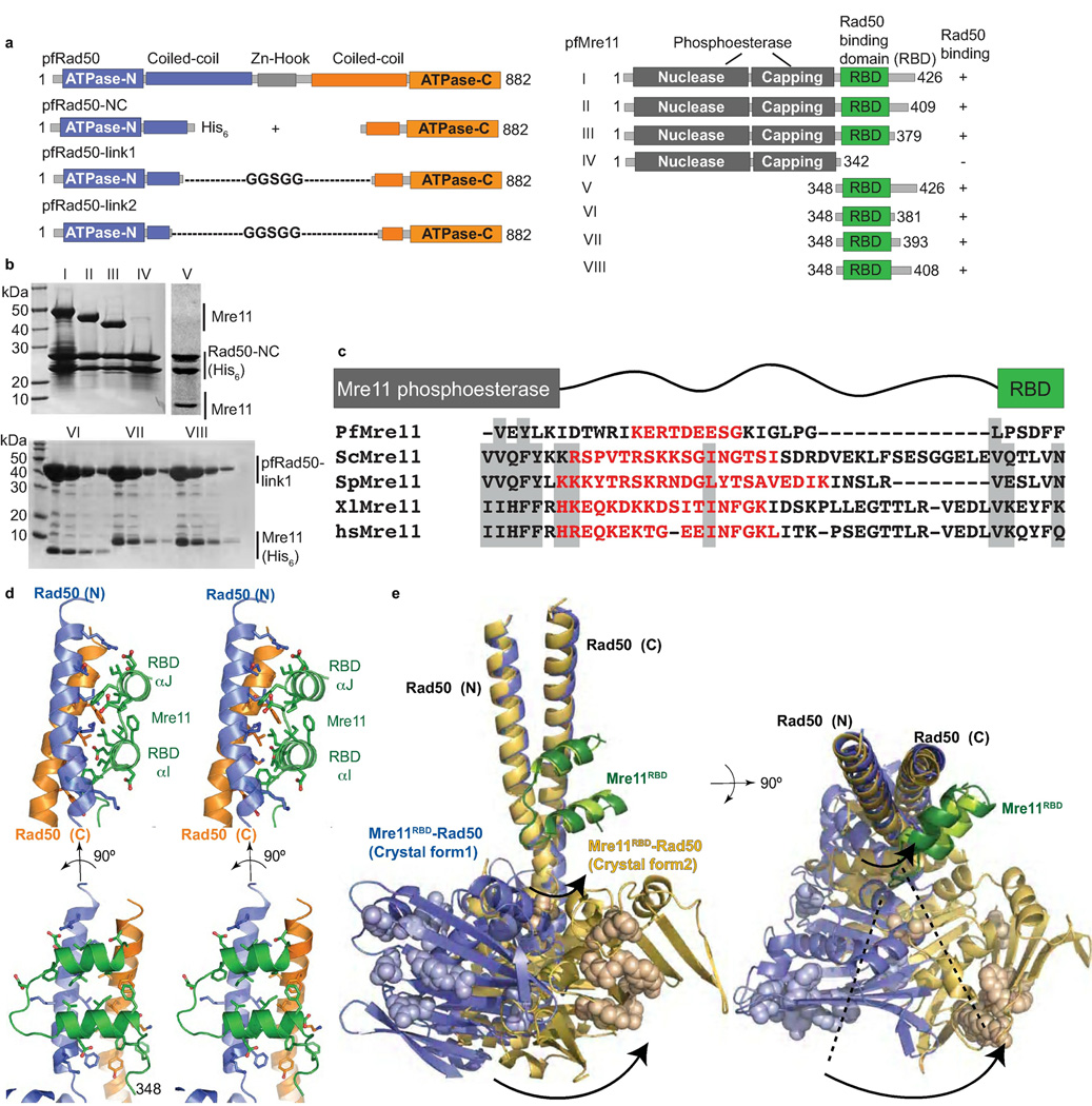
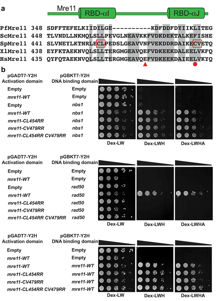
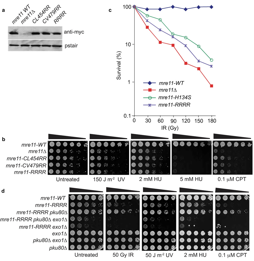
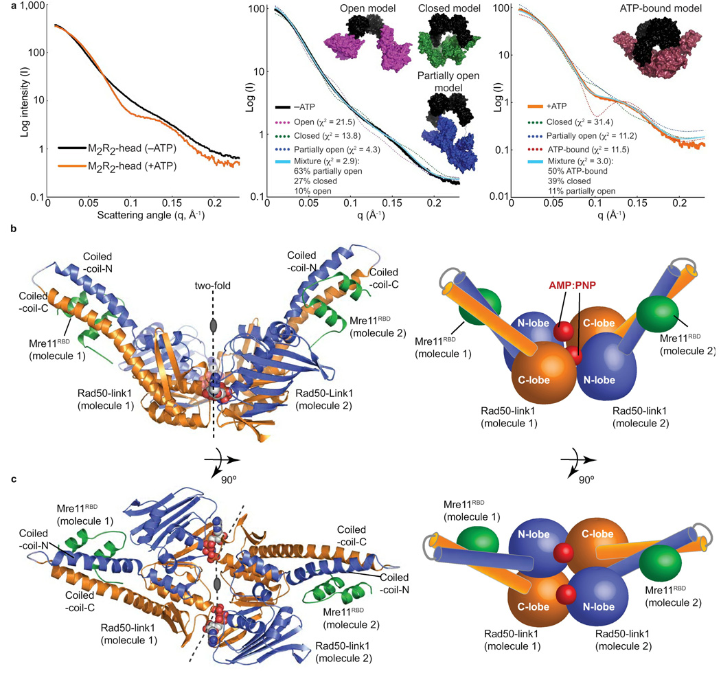
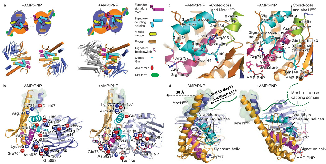
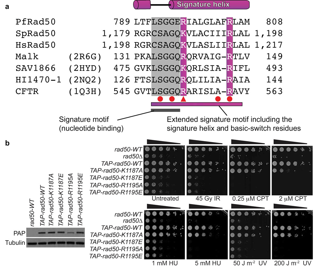
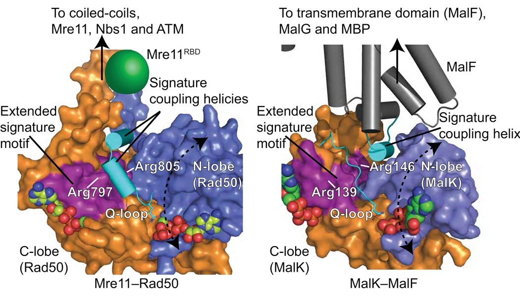
The Rad50 ABC-ATPase complex with Mre11 nuclease is essential for dsDNA break repair, telomere maintenance and ataxia telangiectasia-mutated kinase checkpoint signaling. How Rad50 affects Mre11 functions and how ABC-ATPases communicate nucleotide binding and ligand states across long distances and among protein partners are questions that have remained obscure. Here, structures of Mre11-Rad50 complexes define the Mre11 2-helix Rad50 binding domain (RBD) that forms a four-helix interface with Rad50 coiled coils adjoining the ATPase core. Newly identified effector and basic-switch helix motifs extend the ABC-ATPase signature motif to link ATP-driven Rad50 movements to coiled coils binding Mre11, implying an ~30-Å pull on the linker to the nuclease domain. Both RBD and basic-switch mutations cause clastogen sensitivity. Our new results characterize flexible ATP-dependent Mre11 regulation, defects in cancer-linked RBD mutations, conserved superfamily basic switches and motifs effecting ATP-driven conformational change, and they provide a unified comprehension of ABC-ATPase activities.


Figures

Figure 1
Figure 1. The Mre11RBD–Rad50 interface
(a) pfRad50 and pfMre11 construct schematics for domain mapping and crystallizations. pfRad50-link constructs contain Gly-Ser repeat sequences to intramolecularly link Rad50 N- and C-lobes. (b) Mapping of Mre11 RBD. Top: His6-tagged Rad50 was co-expressed with Mre11 variants (I–V) shown in (a). Bottom: His6-tagged Mre11 variants (V–VIII) were co-expressed with untagged pfRad50-link1. Minimal pfMre11 polypeptide (Mre11RBD, residues 348–381) interacts with Rad50-link1. (c) Sequence alignment of the linker region connecting the Mre11 RBD to the nuclease capping domain in pfMre11, S. cerevisiae (scMre11), S. pombe (spMre11), Xenopus laevis (xlMre11) and human (hsMre11). Shaded regions show well-conserved residues. Disordered residues are shown in red as seen in pfMre11 crystal structures or as predicted by Disopred2. (d) Mre11RBD–Rad50 interface, shown in orthogonal stereo views. The hydrophobic Mre11RBD–Rad50 interaction core is augmented by four flanking complementary salt bridge interactions with acidic residues from Mre11 RBD interacting with 4 positively-charged Rad50 surface residues. (e) Superimposition of two nucleotide-free Mre11RBD–Rad50 crystal forms. The core Mre11RBD–Rad50 interface is maintained, but a ~35° rotation about the base of the Rad50 coiled-coil identifies a flexible linkage to Rad50 ATPase domains. Residues equivalent to those mutated in Rad50S yeast phenotypes are shown in space fill representations.
Figure 2
Figure 2. A conserved interface links eukaryotic Mre11 and Rad50
(a) Multiple sequence alignment of Mre11 RBD from pfMre11, scMre11, spMre11, xlMre11 and hsMre11. Shaded regions show well-conserved residues. CL→RR and CV→RR mark hydrophobic to charged surface substitutions introduced into S. pombe Mre11 RBD. HsMre11 mutations identified in somatic colorectal cancers are highlighted (solid red circle, point mutation; triangle, truncation). (b) S. pombe Mre11 RBD variant interactions with Rad50, Nbs1 and the Mre11 homodimeric interaction analyzed by two-hybrid. Growth on Dex-WL plates (minimal glucose media lacking tryptophan and leucine) indicates the reporter strain transformed with plasmids pGADT7 (Gal4 activating domain) and pGBKT7 (Gal4 DNA binding domain) fused to the respective proteins. Growth on Dex-LWH (less stringent; lacking histidine) and Dex-LWHA (more stringent; lacking histidine and adenine) indicates a positive two-hybrid interaction. We have previously shown that pGBKT7-mre11-WT alone does not autoactivate and this was not repeated here. Mre11 RBD mutants fail to interact with Rad50 yet retain homodimerization and Nbs1 interactions. Strains used are detailed in Supplementary Table 1.
Figure 3
Figure 3. The Mre11–Rad50 interaction interface coordinates DSBR in S. pombe
(a) Expression levels of myc-tagged Mre11 variants. (b) Mre11 RBD variant UV, HU, and CPT genotoxin sensitivity. Mre11–Rad50 interaction interface disruption causes clastogen sensitivity. Five-fold serial dilutions of cells on rich media plates were photographed following 2–3 days at 30°C. (c) Mre11 RBD variants are IR sensitive. This plot is representative of two independent experiments (Supplementary Fig. 2). (d) The IR, UV, HU, and CPT survival defects of mre11-RRRR are suppressed by Ku80 elimination. This rescue depends on Exo1. Strains used are detailed in Supplementary Table 1.
Figure 4
Figure 4. The M2R2-head assembly
(a) SAXS analysis of the M2R2-head reveals a transition from a conformationally flexible, open complex to a globular, closed complex upon ATP binding. Left: experimental SAXS curves of the M2R2-head without (−ATP) and with (+ATP) nucleotide. Fits of single and MES models of M2R2-heads to −ATP (middle panel) and +ATP (right panel) data. Models are shown as surfaces with Mre11 core dimer colored black and Rad50 domains with attached Mre11 RBD colored for open (magenta), partially open (blue), closed (green) and ATP-bound (red) conformations. Fits to the experimental data are shown for single models (dashed line) and the MES ensemble (cyan line) with quality of fit shown by χ2. (b) Mre11RBD–Rad50-link1–AMP:PNP–Mg2+ complex architecture. Left: Mre11RBD (green) binds to Rad50 coiled-coil base. Right: Schematic of the structure. (c) Orthogonal views of the complex as in (b). See Table 1 for data processing and refinement statistics.
Figure 5
Figure 5. Rad50 nucleotide-binding induced conformational changes
(a) Nucleotide free (−AMP:PNP) and bound (AMP:PNP) Rad50 conformations. Nucleotide binding is coordinated by the signature motif, Q-loop, P-loop, and induces a ~35° subdomain rotation. Rad50 N-lobe rotation drives the π-helix wedge into the signature coupling helices, dramatically altering signature coupling helix conformation relative to ATPase subdomain interactions. Motifs are colored as in key. (b) Twenty salt bridge switches rearrange upon nucleotide binding and coordinate domain rotations, see Supplementary Movie 1. Blue (positive) and red (negative) circles highlight charged residues. The Mre11 RBD is highlighted by a green surface representation of Rad50 residues involved in the interface. (c) Signature helix Arg797 and Arg805 rearrangements link nucleotide binding with domain rotations, conformational change of the signature-coupling helices and Q-loop, and motions in the Rad50 coiled-coils (see Supplementary Movie 2). The Mre11 RBD is highlighted as in (b). (d) Nucleotide-binding induced Rad50 ATPase C-lobe rotation relative to the N-lobe drives coiled-coil repositioning to impact bound Mre11 RBD, highlighted as in (b). The Rad50 domain rotation is transduced through coiled-coil repositioning (see Supplementary Movie 3), into a linear pull on the linker between the Mre11 RBD and nuclease capping domain as depicted by dashed arrows.
Figure 6
Figure 6. ABC-ATPase superfamily conserved basic-switch residues in Rad50 coordinate DSBR in S. pombe
(a) Rad50 sequence alignment with ABC transporters shows the extended signature motif, with well-conserved residues shaded. Red circles (point mutations) and triangles (truncations) denote cystic fibrosis causing CFTR mutations. (b) S. pombe Rad50 basic-switch variants on the signature helix are defective for DSBR. Left: Expression levels of TAP-tagged Rad50 variants, as probed by PAP antibody. Right: IR, CPT, HU and UV sensitivity of Rad50 basic-switch variants. Five-fold serial dilutions of cells on rich media plates were photographed following 2–3 days at 30°C. Strains used are detailed in Supplementary Table 1.
Figure 7
Figure 7. Topologically equivalent signature helices connect nucleotide binding to conformational changes in the ABC-ATPase superfamily
Rad50 ABC-ATPase molecular surface with attached coiled-coil and mapped Mre11 RBD compared to MalK ABC-ATPase with interacting MalF transmembrane protein. The extended signature helix (purple) and signature coupling helices or helix (cyan) connect nucleotide binding to movements of attached functional domains and proteins.

References

    1. Paull TT, Gellert M. Nbs1 potentiates ATP-driven DNA unwinding and endonuclease cleavage by the Mre11/Rad50 complex. Genes Dev. 1999;13:1276–1288. - PMC - PubMed
    1. Trujillo KM, Sung P. DNA structure-specific nuclease activities in the Saccharomyces cerevisiae Rad50-Mre11 complex. J. Biol. Chem. 2001;276:35458–35464. - PubMed
    1. Williams GJ, Lees-Miller SP, Tainer JA. Mre11-Rad50-Nbs1 conformations and the control of sensing, signaling, and effector responses at DNA double-strand breaks. DNA Repair (Amst) 2010;9:1299–1306. - PMC - PubMed
    1. Williams RS, Williams JS, Tainer JA. Mre11-Rad50-Nbs1 is a keystone complex connecting DNA repair machinery, double-strand break signaling, and the chromatin template. Biochem. Cell Biol. 2007;85:509–520. - PubMed
    1. Carney JP, et al. The hMre11/hRad50 protein complex and Nijmegen breakage syndrome: linkage of double-strand break repair to the cellular DNA damage response. Cell. 1998;93:477–486. - PubMed

Publication types

MeSH terms