. 2011 May;14(5):578-86.
doi: 10.1038/nn.2798. Epub 2011 Mar 27.

Deletion of a remote enhancer near ATOH7 disrupts retinal neurogenesis, causing NCRNA disease

Affiliations

Deletion of a remote enhancer near ATOH7 disrupts retinal neurogenesis, causing NCRNA disease

Noor M Ghiasvand et al. Nat Neurosci. 2011 May.

Abstract

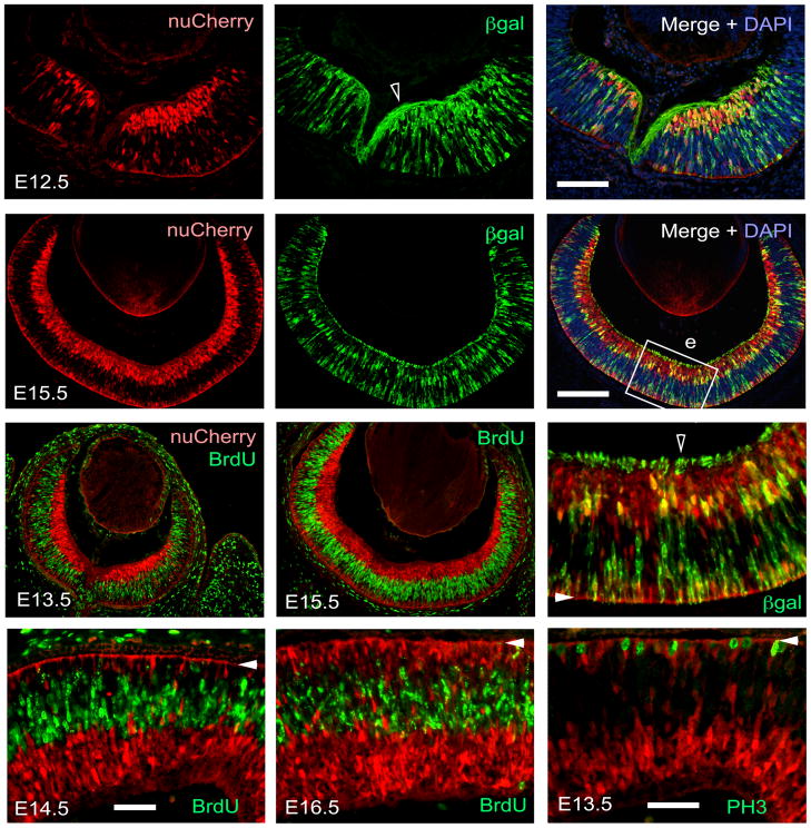
Individuals with nonsyndromic congenital retinal nonattachment (NCRNA) are totally blind from birth. The disease afflicts ∼1% of Kurdish people living in a group of neighboring villages in North Khorasan, Iran. We found that NCRNA is caused by a 6,523-bp deletion that spans a remote cis regulatory element 20 kb upstream from ATOH7 (Math5), a bHLH transcription factor gene that is required for retinal ganglion cell (RGC) and optic nerve development. In humans, the absence of RGCs stimulates massive neovascular growth of fetal blood vessels in the vitreous and early retinal detachment. The remote ATOH7 element appears to act as a secondary or 'shadow' transcriptional enhancer. It has minimal sequence similarity to the primary enhancer, which is close to the ATOH7 promoter, but drives transgene expression with an identical spatiotemporal pattern in the mouse retina. The human transgene also functions appropriately in zebrafish, reflecting deep evolutionary conservation. These dual enhancers may reinforce ATOH7 expression during early critical stages of eye development when retinal neurogenesis is initiated.


Figures

Figure 1
Figure 1
NCRNA disease. AF. Eye photographs from NCRNA family. A. 49-yr old female B. 18-yr old male C. 14-yr old female D. 8-yr old male. Retinal detachments are evident as leukocoria (white pupils) in each case. The NCRNA patients eventually develop dense gray posterior corneal opacities, consistent with chronic blood staining (A, B, patient’s right eye in C). They typically exhibit nystagmus and strabismus (esotropia, D). The pupils are round, but do not react to light. E–F. Closer views showing leukocoria in young patients prior to corneal opacification. E. 5-yr old male (left eye). F. 3-yr old male (left eye). The detached retina and fibrovascular mass are visible behind the clear lens (arrow). G. Khorasani pedigree showing autosomal recessive inheritance of the disease over nine generations, with index cases used for mutation screening (red boxes).
Figure 2
Figure 2
Anatomical findings in NCRNA. A–C. Orbital magnetic resonance images (MRI) of an 18-yr old male patient, showing absence or severe atrophy of the optic nerves and chiasm (red arrowheads), and bilateral detached retinas with vitreous consolidation (bright signal, blue arrowheads). A. Axial (TR/TE = 780/15 ms, 3 mm slices) and B. Oblique sagittal (right eye, TR/TE = 730/15 ms, 2 mm slice) T1-weighted SE views, with fat suppression and gadolinium contrast. C. Coronal T1-weighted SE views (TR/TE = 780/15, 5 mm slices) with no contrast or fat suppression, shown from anterior (left) to posterior (right). D. Matched coronal images from a normal 15-yr old female. T1-weighted SE views (TR/TE = 550/11 ms, 3 mm slices). The vitreae are clear (left panel, dark signal, blue arrowhead) and the optic nerves are prominent (middle panels, red arrowheads). The optic chiasm is visible above the suprasellar cistern and pituitary (right panel, red arrowhead). E. Matched axial images from the normal subject (TR/TE = 750/11 ms, 3 mm slices). F,G. Enlarged coronal views of NCRNA and normal subjects (left orbits, boxed areas in panels C and D). The red arrowhead shows the normal optic nerve. The bright T1W signal outlining the optic nerve, extraocular muscles (sr, lr, ir, mr, so, lps) and blood vessels (sov) originates from retrobulbar fat. 3v, third ventricle; cc, corpus callosum; cis, suprasellar cistern; eth, ethmoid sinus; hyp, hypothalamus; ica, internal carotid artery; io, inferior oblique; ir, inferior rectus; la, levator aponeurosis; lg, lacrimal gland; lps, levator palpebrae superioris; lr, lateral rectus; lv, lateral ventricle; max, maxillary sinus; mr, medial rectus; nph, nasopharynx; oa, ophthalmic artery; oc, optic canal; on, optic nerve; pit, pituitary gland; po, pons; ps, pituitary stalk; so, superior oblique; sov, superior orbital vein; sph, sphenoid sinus; sr, superior rectus; st, sella turcica; tem, temporal lobe; vit, vitreous; x, optic chiasm.
Figure 2
Figure 2
Anatomical findings in NCRNA. A–C. Orbital magnetic resonance images (MRI) of an 18-yr old male patient, showing absence or severe atrophy of the optic nerves and chiasm (red arrowheads), and bilateral detached retinas with vitreous consolidation (bright signal, blue arrowheads). A. Axial (TR/TE = 780/15 ms, 3 mm slices) and B. Oblique sagittal (right eye, TR/TE = 730/15 ms, 2 mm slice) T1-weighted SE views, with fat suppression and gadolinium contrast. C. Coronal T1-weighted SE views (TR/TE = 780/15, 5 mm slices) with no contrast or fat suppression, shown from anterior (left) to posterior (right). D. Matched coronal images from a normal 15-yr old female. T1-weighted SE views (TR/TE = 550/11 ms, 3 mm slices). The vitreae are clear (left panel, dark signal, blue arrowhead) and the optic nerves are prominent (middle panels, red arrowheads). The optic chiasm is visible above the suprasellar cistern and pituitary (right panel, red arrowhead). E. Matched axial images from the normal subject (TR/TE = 750/11 ms, 3 mm slices). F,G. Enlarged coronal views of NCRNA and normal subjects (left orbits, boxed areas in panels C and D). The red arrowhead shows the normal optic nerve. The bright T1W signal outlining the optic nerve, extraocular muscles (sr, lr, ir, mr, so, lps) and blood vessels (sov) originates from retrobulbar fat. 3v, third ventricle; cc, corpus callosum; cis, suprasellar cistern; eth, ethmoid sinus; hyp, hypothalamus; ica, internal carotid artery; io, inferior oblique; ir, inferior rectus; la, levator aponeurosis; lg, lacrimal gland; lps, levator palpebrae superioris; lr, lateral rectus; lv, lateral ventricle; max, maxillary sinus; mr, medial rectus; nph, nasopharynx; oa, ophthalmic artery; oc, optic canal; on, optic nerve; pit, pituitary gland; po, pons; ps, pituitary stalk; so, superior oblique; sov, superior orbital vein; sph, sphenoid sinus; sr, superior rectus; st, sella turcica; tem, temporal lobe; vit, vitreous; x, optic chiasm.
Figure 3
Figure 3
Homozygous deletion of 5′ ATOH7 genomic sequences in NCRNA patients. A. The 1639 kb critical region on chromosome 10q21 spans 14 positional candidate genes. The segment between PBLD to MYPN (blue bracket) is expanded below. B. Genomic PCRs showing deletion of four adjacent amplicons in an NCRNA patient. Two additional amplicons were also missing (Supplementary Table 3). C. Map of 74 kb intergenic region surrounding ATOH7 (Chr10:69,714,052–69,640,048) modified from the UCSC browser (NCBI36/hg18, Mar 2006 assembly), showing the terminal exons of flanking genes; KRTψ (human-specific keratin-18 pseudogene); vertebrate and mammalian evolutionary conservation tracks (PhastCons); interspersed repetitive elements; PCR amplicons (red) used to compare homozygous mutant (rr) and wild-type (RR) DNA samples; and the 6.5 kb NCRNA deletion. Orthologous vertebrate sequences are indicated in the vertebrate MultiZ alignment. In the nontherian genomes, similarity between KRTψ and unlinked keratin loci gives a false positive signal of homologous evolution. The corresponding mouse Pbld-Mypn intergenic segment is 42.5 kb. Two SNPs (single nucleotide polymorphisms) associated with optic disc area in human genome-wide studies, are indicated for comparison (rs1900004, rs3858145). The deletion removes a cluster of CNEs (conserved noncoding elements) ~20 kb upstream of ATOH7.
Figure 4
Figure 4
Endpoints of the NCRNA deletion. A. Triplex PCR genotypes showing transmission of the mutation in a small family. B. Sequence chromatograms from PCR products showing the distal and proximal breakpoints in wild-type DNA, with 5 bp microhomology (double arrow), and the deletion junction in a DNA from a blind individual.
Figure 5
Figure 5
The NCRNA mutation deletes an ATOH7 retinal enhancer. A. Map of the 30kb upstream region (Chr10:69,690,000–69,660,000) modified from the UCSC browser (NCBI36/hg18, Mar 2006 assembly), showing mammalian basewise sequence conservation and the NCRNA deletion. The 3034-BGnCherry transgene contains 3.8 kb human genomic DNA, which spans the three deleted CNEs and regulates expression from the minimal human β-globin promoter. The location of the Math5-GFP transgene is shown for comparison. This reporter contains the endogenous mouse Atoh7 promoter, 0.3 kb 5′UTR and 1.8 kb upstream genomic DNA. B–E. Brightfield (B,C) and fluorescence (D,E) images show eye-specific nuCherry expression in a pigmented E13.5 transgenic founder. Tg, transgenic.
Figure 6
Figure 6
Activity of the remote ATOH7 retinal enhancer in 3034-BGnCherry mice. Double transgenic 3034-BGnCherry; Math5-lacZ/+ embryos were exposed to BrdU, harvested between E12 and E16, cryosectioned, and immunostained for nuCherry (red) and β-galactosidase, BrdU or phosphohistone H3 (green). A,B,E. The nuclear 3034- BGnCherry and cytoplasmic Math5-lacZ patterns are essentially overlapping, with >85% cell concordance (not shown). However, the subcellular localizations differ, with accumulation of βgal in RGC axons at the inner retinal surface and nascent optic nerve (open arrowheads). C,D,F–H. The 3034-BGnCherry transgene is expressed in post-mitotic retinal cells, with no overlap between nuCherry and the mitotic markers BrdU (S-phase) or PH3 (M-phase). Closed arrowheads mark the apical (sclerad) side in E–H. Scale bars: 40 μm in A; 20 μm in D; and 10 μm in E–H.
Figure 7
Figure 7
Remote and primary ATOH7 enhancers have similar activity in the developing mouse retina. Coexpression of nuCherry and GFP transgenes. A–B. Double transgenic 3034-BGnCherry; Math5-GFP embryos immunostained for both reporters. Low (A) and confocal high (B) magnification views of E11.5 to E16.5 retinal sections. The onset and expression patterns of the transgenes are essentially overlapping. C. Two-dimensional flow cytometric analysis of dissociated E14.5 retinal cells from 3034-BGnCherry × Math5-GFP littermate embryos carrying one, neither or both transgenes. The percentage of cells in each quadrant is indicated in the contour plots. The concordance between nuCherry and GFP fluorescence is high. Double-positive cells represent ~40% of the neural retina in 3034-BGnCherry; Math5-GFP embryos (upper right). D. Dissociated cells from an E14.5 double transgenic embryo immunostained for nuCherry and GFP. Arrowheads in B mark the apical side. Scale bars: 40 μm in A; 20 μm in B; and 10 μm in D.
Figure 8
Figure 8
The human remote ATOH7 enhancer functions in developing zebrafish. A,B. Lateral and dorsal views of live embryos showing specific retinal expression of the 3034-BGnCherry transgene in progenitor cells at 72 hpf development. C–E. Transverse sections of immunostained 3034-BGnCherry; ath5:GFP embryos showing colocalized expression of nuCherry and GFP in the retina at 48, 72 and 128 hpf, with perdurance of reporter proteins in RGCs. The cytoplasmic GFP also labels RGC axons in the optic nerves, chiasm and tracts projecting to the thalamus and tectum. g, ganglion cell layer; hb, habenula; i, inner nuclear layer; o, outer nuclear layer; on, optic nerve; rtt, retinotectal tract; tel, telencephalon; th, thalamus; hpf, hours post fertilization. Scale bars, 50 μm.

References

    1. Livesey FJ, Cepko CL. Vertebrate neural cell-fate determination: lessons from the retina. Nat Rev Neurosci. 2001;2:109–118. - PubMed
    1. West H, Richardson WD, Fruttiger M. Stabilization of the retinal vascular network by reciprocal feedback between blood vessels and astrocytes. Development. 2005;132:1855–1862. - PubMed
    1. Brown NL, et al. Math5 encodes a murine basic helix-loop-helix transcription factor expressed during early stages of retinal neurogenesis. Development. 1998;125:4821–4833. - PubMed
    1. Brzezinski JA, Glaser T. Math5 establishes retinal ganglion cell competence in postmitotic progenitor cells. Invest Ophthalmol Vis Sci. 2004;45:3422. E-abstract.
    1. Brown NL, Patel S, Brzezinski J, Glaser T. Math5 is required for retinal ganglion cell and optic nerve formation. Development. 2001;128:2497–2508. - PMC - PubMed

Publication types

MeSH terms

Substances