. 2011 May;43(5):470-5.
doi: 10.1038/ng.796. Epub 2011 Mar 27.

Mutant nucleophosmin and cooperating pathways drive leukemia initiation and progression in mice

Affiliations

Mutant nucleophosmin and cooperating pathways drive leukemia initiation and progression in mice

George S Vassiliou et al. Nat Genet. 2011 May.

Abstract

Acute myeloid leukemia (AML) is a molecularly diverse malignancy with a poor prognosis whose largest subgroup is characterized by somatic mutations in NPM1, which encodes nucleophosmin. These mutations, termed NPM1c, result in cytoplasmic dislocation of nucleophosmin and are associated with distinctive transcriptional signatures, yet their role in leukemogenesis remains obscure. Here we report that activation of a humanized Npm1c knock-in allele in mouse hemopoietic stem cells causes Hox gene overexpression, enhanced self renewal and expanded myelopoiesis. One third of mice developed delayed-onset AML, suggesting a requirement for cooperating mutations. We identified such mutations using a Sleeping Beauty transposon, which caused rapid-onset AML in 80% of mice with Npm1c, associated with mutually exclusive integrations in Csf2, Flt3 or Rasgrp1 in 55 of 70 leukemias. We also identified recurrent integrations in known and newly discovered leukemia genes including Nf1, Bach2, Dleu2 and Nup98. Our results provide new pathogenetic insights and identify possible therapeutic targets in NPM1c+ AML.


Figures

Figure 1
Figure 1. Conditional mouse model of type A NPM1c mutation
a, N-terminal GFP-fusions of type A human (NPM1cA) and “humanized” mouse (Npm1cA) mutants show identical sub-cellular distribution. b, The conditional Npm1flox-cA allele interferes minimally with the native locus and converts to mutant Npm1cA allele by Cre (humanized mutant exon 11* in red; mouse exon 11 is homologous to human exon 12). c-d, Npm1cA RNA and protein detected in Post-Cre ES cells by RT-PCR (c) and Western Blot (d) respectively. e, Universal lethality of Npm1flox-cA/+; Stella-Cre+ (** p=0.0001) versus mendelian ratios for Npm1flox-cA/+; Mx1-Cre+ mice. P, PstI site; PuΔTK, Puro-Delta-TK cassette; K562-Npm1, K562 cells transfected with Npm1cA or Npm1WT cDNA; RT, Reverse Transcriptase, RT-PCR primers: R1, forward; R2, Npm1WT reverse; R3, Npm1cA reverse (red).
Figure 2
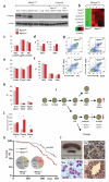
Figure 2. Hematopoietic changes and incidence of AML in Npm1cA/+ mice
a, Hemopoietic expression of Npm1cA protein in Npm1cA/+ mice. b, Hox-overexpression in Npm1cA/+ vs Npm1WT lineage negative hemopoietic progenitors. c,d, No significant differences in white cell (WCC), hemoglobin (Hb) or platelet counts (Plts), but higher mean red cell (MCV) and platelet (MPV) volumes in Npm1cA/+ mice. e,f, No differences in marrow stem and progenitor cell compartment sizes. g,h, Expansion of mature myeloid (Gr1+/Mac1+) and reduction in late (B220+/CD19+) B-cells in Npm1cA/+ vs Npm1WT marrow. i, Summary of hemopoietic changes in Npm1cA/+ mice. j, Increased serial re-plating of Npm1cA/+ myeloid progenitors. k, Decreased survival of Npm1cA/+ mice due to excess AML. l, Example of AML showing splenomegaly due to infiltration with myeloperoxidase-positive blasts (Sp), also infiltrating the liver (Li) and blood (Bld). Error bars show the standard error of the mean; *p<0.05; B,T, B or T-cell leukemia/lymphoma; Non-Hem, Non-hematological.
Figure 3
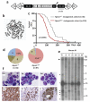
Figure 3. Npm1cA and the GrOnc transposon synergize to cause AML
a, The GrOnc transposon carrying gene-activating and inactivating elements flanked by repeats for the Sleeping Beauty (SB) and PiggyBac (PB). b, Blood-metaphase FISH showing the genomic location of the GrOnc donor locus on chromosome 19. c, Acceleration of leukemogenesis in Npm1cA/+ compared to Npm1+/+ mutagenised mice. d, Marked increase in the proportion of AMLs and absence of T cell tumors in Npm1cA/+ compared to Npm1+/+ mice. e, Southern blot showing clonal transposon integrations in mouse leukemias. Endogenous En2 (arrowhead) and GrOnc donor locus concatamer (dots) bands indicated. f, Morphology and immunohistochemistry (anti-myeloperoxidase) from an Npm1cA/+ AML. Gr1.4 LTR, Graffi1.4 MuLV long terminal repeat; SD, Lun splice donor; En2-SA and βA-SA, Engrailed 2 and Carp β-actin splice acceptor; pA, adenovirus polyadenylation signal; *p<0.00001; B, T and U, B-cell, T-cell and undifferentiated leukemia/lymphoma; Bld, blood; Sp, spleen; Sar, myeloid sarcoma; Ki, kidney; Li, liver.
Figure 4
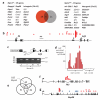
Figure 4. Common integration sites in transposon-derived leukemias
a, Common integration sites in Npm1cA/+ and Npm1+/+mice show some overlap (regular font) but are mostly different (bold). b-d, Directional GrOnc integrations at the Csf2 locus identified in 42 of 70 AMLs, were associated with the formation of two LunSD-Csf2 fusion mRNA splice variants (sv1 and sv2) and marked overexpression of Csf2 mRNA (note break in Y axis). e, GrOnc integrations in three individual myeloid blast colonies from one of these AMLs shared only 2 insertions, involving Csf2 and the myeloid oncogene Myst4(Moz) suggesting that these were “driver” insertions. e, Directional activating integrations in intron 9 of Flt3. g, Bi-directional integrations in Nup98.
Figure 5
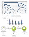
Figure 5. A model for Npm1cA/+-driven leukemogenesis
a, Co-occurrence table of CIS genes in 70 NPm1cA/+ AMLs reveals mutually exclusive insertions in Csf2, Flt3, Rasgrp1 and Kras. b, Persistence of Hox overexpression in Npm1cA/+ vs Npm1+/+ AMLs indicating that this effect of Npm1cA persists in leukemic cells(* p≤0.03). c. Model for Npm1cA - driven leukemogenesis supported by our data: isolated Npm1cA increases self renewal but leukemic transformation only occurs upon activation of defined proliferative pathways (Type I mutation), facilitated by a permissive transcription factor (TF) mutation. Error bars show the standard error of the mean. Localization of Npm1 protein indicated in green.

References

    1. Falini B, et al. Cytoplasmic nucleophosmin in acute myelogenous leukemia with a normal karyotype. N Engl J Med. 2005;352:254–266. - PubMed
    1. Alcalay M, et al. Acute myeloid leukemia bearing cytoplasmic nucleophosmin (NPMc+ AML) shows a distinct gene expression profile characterized by up-regulation of genes involved in stem-cell maintenance. Blood. 2005;106:899–902. doi:2005-02-0560 [pii] 10.1182/blood-2005-02-0560. - PubMed
    1. Dupuy AJ, Akagi K, Largaespada DA, Copeland NG, Jenkins NA. Mammalian mutagenesis using a highly mobile somatic Sleeping Beauty transposon system. Nature. 2005;436:221–226. - PubMed
    1. Collier LS, Carlson CM, Ravimohan S, Dupuy AJ, Largaespada DA. Cancer gene discovery in solid tumours using transposon-based somatic mutagenesis in the mouse. Nature. 2005;436:272–276. doi:nature03681 [pii] 10.1038/nature03681. - PubMed
    1. Okuwaki M. The structure and functions of NPM1/Nucleophsmin/B23, a multifunctional nucleolar acidic protein. J Biochem. 2008;143:441–448. doi:mvm222 [pii] 10.1093/jb/mvm222. - PubMed

Publication types

MeSH terms