

Promoting effects of isobavachin on neurogenesis of mouse embryonic stem cells were associated with protein prenylation
- PMID: 21441946
- PMCID: PMC4001985
- DOI: 10.1038/aps.2011.5
Promoting effects of isobavachin on neurogenesis of mouse embryonic stem cells were associated with protein prenylation
Abstract
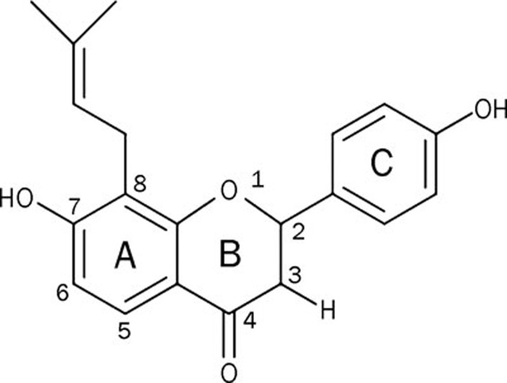
Aim: Some small molecules can induce mouse embryonic stem (ES) cells to differentiate into neuronal cells. Here, we explored the effect of isobavachin (IBA), a compound with a prenyl group at position 8 of ring A, on promoting neuronal differentiation and the potential role of its protein prenylation.
Methods: The hanging drop method was employed for embryonic body (EB) formation to mimic embryo development in vivo. The EBs were treated with IBA at a final concentration of 10(-7) mol/L from EB stage (d 4) to d 8+10. Geranylgeranyltransferase I inhibitor GGTI-298 was subsequently used to disrupt protein prenylation. Neuronal subtypes, including neurons and astrocytes, were observed by fluorescence microscopy. Gene and protein expression levels were detected using RT-PCR and Western blot analysis, respectively.
Results: With IBA treatment, nestin was highly expressed in the neural progenitors generated from EBs (d 4, d 8+0). EBs then further differentiated into neurons (marked by β-tubulin III) and astrocytes (marked by GFAP), which were both up-regulated in a time-dependent manner on d 8+5 and d 8+10. Co-treatment with GGTI-298 selectively abolished the IBA-induced neuronal differentiation. Moreover, in the MAPK pathway, p38 and JNK phosphorylation were down-regulated, while ERK phosphorylation was up-regulated after IBA treatment at different neuronal differentiation passages.
Conclusion: IBA can facilitate mouse ES cells differentiating into neuronal cells. The mechanism involved protein prenylation and, subsequently, phos-ERK activation and the phos-p38 off pathway.
Figures


Similar articles
-
Enhanced co-expression of beta-tubulin III and choline acetyltransferase in neurons from mouse embryonic stem cells promoted by icaritin in an estrogen receptor-independent manner.Chem Biol Interact. 2009 May 15;179(2-3):375-85. doi: 10.1016/j.cbi.2008.12.007. Epub 2008 Dec 16. Chem Biol Interact. 2009. PMID: 19135036
-
[Directed differentiation of mouse embryonic stem cells into neuronal cells induced by icaritin in vitro].Zhejiang Da Xue Xue Bao Yi Xue Ban. 2007 May;36(3):217-23. doi: 10.3785/j.issn.1008-9292.2007.03.002. Zhejiang Da Xue Xue Bao Yi Xue Ban. 2007. PMID: 17571302 Chinese.
-
2,4-Dinitrophenol induces neural differentiation of murine embryonic stem cells.Stem Cell Res. 2013 Nov;11(3):1407-16. doi: 10.1016/j.scr.2013.09.016. Epub 2013 Oct 6. Stem Cell Res. 2013. PMID: 24148244
-
[Phenotype-based primary screening for drugs promoting neuronal subtype differentiation in embryonic stem cells with light microscope].Zhejiang Da Xue Xue Bao Yi Xue Ban. 2012 Jul;41(4):373-80. Zhejiang Da Xue Xue Bao Yi Xue Ban. 2012. PMID: 22927071 Chinese.
-
Distinct contributions of JNK and p38 to chromium cytotoxicity and inhibition of murine embryonic stem cell differentiation.Environ Health Perspect. 2009 Jul;117(7):1124-30. doi: 10.1289/ehp.0800157. Epub 2009 Apr 3. Environ Health Perspect. 2009. PMID: 19654923 Free PMC article.
Cited by
-
A Flavonoid Compound Promotes Neuronal Differentiation of Embryonic Stem Cells via PPAR-β Modulating Mitochondrial Energy Metabolism.PLoS One. 2016 Jun 17;11(6):e0157747. doi: 10.1371/journal.pone.0157747. eCollection 2016. PLoS One. 2016. PMID: 27315062 Free PMC article.
-
Flavonoids in Combination with Stem Cells for the Treatment of Neurological Disorders.Neurochem Res. 2023 Nov;48(11):3270-3282. doi: 10.1007/s11064-023-03986-w. Epub 2023 Jul 18. Neurochem Res. 2023. PMID: 37462837 Review.
-
Investigation on the metabolic characteristics of isobavachin in Psoralea corylifolia L. (Bu-gu-zhi) and its potential inhibition against human cytochrome P450s and UDP-glucuronosyltransferases.J Pharm Pharmacol. 2020 Dec;72(12):1865-1878. doi: 10.1111/jphp.13337. Epub 2020 Aug 4. J Pharm Pharmacol. 2020. PMID: 32750744 Free PMC article.
-
Phenotypic technologies in stem cell biology.Cell Chem Biol. 2021 Mar 18;28(3):257-270. doi: 10.1016/j.chembiol.2021.02.001. Epub 2021 Mar 1. Cell Chem Biol. 2021. PMID: 33651977 Free PMC article. Review.
-
Shen-Jing as a Chinese Medicine Concept Might Be a Counterpart of Stem Cells in Regenerative Medicine.Chin J Integr Med. 2019 Jan;25(1):64-70. doi: 10.1007/s11655-015-2136-z. Epub 2015 Jul 4. Chin J Integr Med. 2019. PMID: 26142336
References
-
- MacLaren RE, Pearson RA. Stem cell therapy and the retina. Eye (Lond) 2007;21:1352–9. - PubMed
-
- Xing F, Fang Z, Qin B, Li Y, Hou J, Chen X. Parthenogenetic embryonic stem cells derived from cryopreserved newborn mouse ovaries: a new approach to autologous stem cell therapy. Fertil Steril. 2009;91:1238–44. - PubMed
-
- Emre N, Coleman R, Ding S. A chemical approach to stem cell biology. Curr Opin Chem Biol. 2007;11:252–8. - PubMed
-
- Trompouki E, Zon LI. Small molecule screen in zebrafish and HSC expansion. Methods Mol Biol. 2010;636:301–16. - PubMed
-
- Xu Y, Shi Y, Ding S. A chemical approach to stem-cell biology and regenerative medicine. Nature. 2008;453:338–44. - PubMed
Publication types
MeSH terms
Substances
LinkOut - more resources
Full Text Sources
Research Materials
Miscellaneous
