Review
. 2011 May;210(3-4):331-45.
doi: 10.1007/s00221-011-2611-5. Epub 2011 Mar 26.

Vestibular control of the head: possible functions of the vestibulocollic reflex

Affiliations
Review

Vestibular control of the head: possible functions of the vestibulocollic reflex

Jay M Goldberg et al. Exp Brain Res. 2011 May.

Abstract

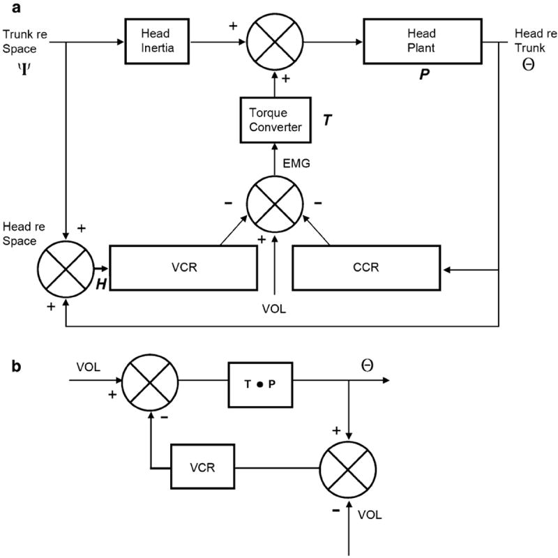
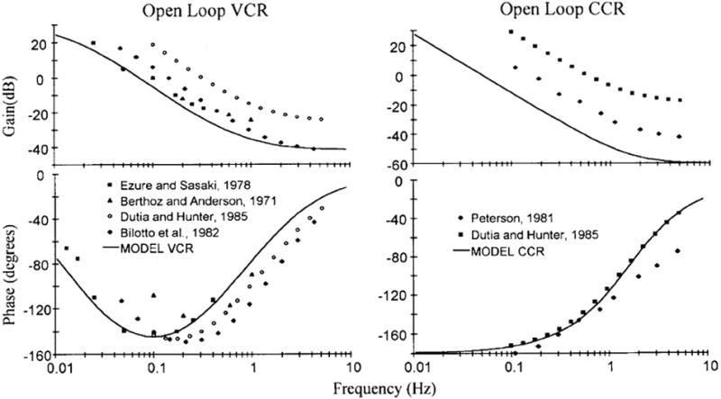
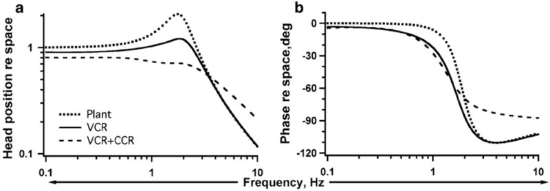
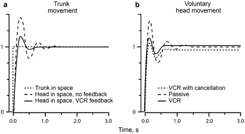
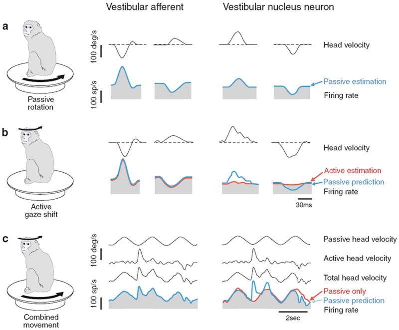
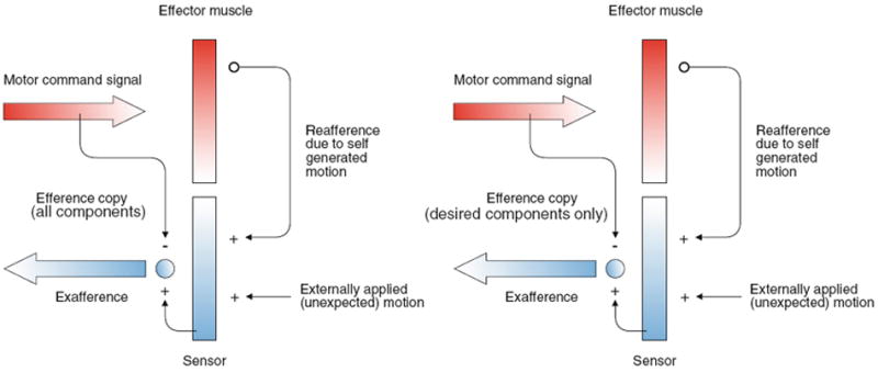
Here, we review the angular vestibulocollic reflex (VCR) focusing on its function during unexpected and voluntary head movements. Theoretically, the VCR could (1) stabilize the head in space during body movements and/or (2) dampen head oscillations that could occur as a result of the head's underdamped mechanics. The reflex appears unaffected when the simplest, trisynaptic VCR pathways are severed. The VCR's efficacy varies across species; in humans and monkeys, head stabilization is ineffective during low-frequency body movements in the yaw plan. While the appearance of head oscillations after the attenuation of semicircular canal function suggests a role in damping, this interpretation is complicated by defects in the vestibular input to other descending motor pathways such as gaze premotor circuits. Since the VCR should oppose head movements, it has been proposed that the reflex is suppressed during voluntary head motion. Consistent with this idea, vestibular-only (VO) neurons, which are possible vestibulocollic neurons, respond vigorously to passive, but not active, head rotations. Although VO neurons project to the spinal cord, their contribution to the VCR remains to be established. VCR cancelation during active head movements could be accomplished by an efference copy signal negating afferent activity related to active motion. Oscillations occurring during active motion could be eliminated by some combination of reflex actions and voluntary motor commands that take into account the head's biomechanics. A direct demonstration of the status of the VCR during active head movements is required to clarify the function of the reflex.


Figures

Fig. 1
Fig. 1
Head movements induced in the cat by electric-shock stimulation of individual ampullary nerves are in the plane of the corresponding canal. Comparison with the directional properties of canals indicates that the evoked head movements are opposite in direction to the head rotations that would excite the individual canals or canal pairs. RAC and LAC right and left anterior canals, RPC and LPC right and left posterior canals, RLC right lateral canal a. Stimulation of individual vertical canals evokes diagonal movements. b Stimulation of pairs of vertical canals produces movements in pitch or roll. c RLC stimulation produces yaw movement to the left. From Suzuki and Cohen 1964
Fig. 2
Fig. 2
Effects of cutting the MLF ipsilateral to a dorsal neck motoneuron from which synaptic potentials were recorded in response to stimulation of contralateral ampullary nerves. ST solitary tract, XII hypoglossal nerve, IO inferior olive, PT pyramidal tract, MLF medial longitudinal fasciculus. Stimulated contralateral ampullary nerve for each trace: Ant anterior canal, Hor horizontal canal, Pos posterior canal. Lesion is shown by the dark area in a. Responses recorded in a complexus motoneuron before (b) and after the cut (c). Stimulus was 50 μA (Hor) and 75 μA (Ant and Pos) before the cut. All stimuli were 75 μA after the cut. Potentials are averages of 50 sweeps. From Wilson and Maeda (1974)
Fig. 3
Fig. 3
Pathways between ipsilateral and contralateral ampullary nerves and neck motoneurons. Inhibitory vestibulocollic neurons and their terminals are shown in black; excitatory vestibulocollic neurons, in white. a Pattern observed in motoneurons innervating dorsal neck muscles, including biventer and complexus. MVST axons travel in the MLF. From Wilson and Maeda (1974). b Pattern observed in motoneurons of a rotator, obliquus capitis caudalis. Axons in circle are in the MVST; those outside the circle are in the LVST. Note that the inhibition from the contralateral anterior canal acts via an inhibitory commissural neuron at segmental levels. From Sugiuchi et al. (2004). A, H, P, anterior, horizontal, and posterior ampullae, VN vestibular nuclei, MN motoneuron, MLF medial longitudinal fasciculus, LVST and MVST lateral and medial vestibulospinal tracts
Fig. 4
Fig. 4
Dynamics of the vestibulocollic reflex (VCR). a EMG responses from a single unit in a dorsal neck muscle. Data are expressed re angular position for responses to different frequencies (Hz) of sinusoidal horizontal rotation. b Bode plot of 5 motor units. From Ezure and Sasaki (1978). c Phase lags of the VCR and of primary vestibular afferents compiled by Bilotto et al. (1982). Multi-unit EMG from dorsal neck muscles. Filled squares VCR phase from one experiment (complexus muscle). Filled triangles average VCR data from Ezure and Sasaki (1978). Open squares data from Berthoz and Anderson (1971). Upper curves: filled circles mean phase of primary afferent population; open circles phase of a single irregular primary afferent from Tomko et al. (1981)
Fig. 5
Fig. 5
Vestibular-evoked myogenic potentials (VEMPs) evoked by a 100-db normal hearing level click delivered to the right ear via earphones. The traces consist of averaged unrectified electromyograms (EMGs) from the sternocleidomastoid muscles (SCMs) on the two sides. A large biphasic (p13-n23) response is seen ipsilateral to the stimulated ear (R SCM) and a smaller response of opposite polarity (n1-p1) is seen contralaterally (L SCM). From Welgampola and Colebatch (2005)
Fig. 6
Fig. 6
a Block diagram of head–neck plant including input from trunk re space (ψ) and output, head re trunk (neck, Θ). Two reflexes (VCR and CCR) sum with voluntary motor commands (VOL) to result in muscle activation (EMG), which is transformed to torques by a low-pass torque converter (T). Head re space (H) is the sum of ψ and Θ. Head inertia (I) and passive plant mechanics (P). Details in the “Appendix”. From Peng et al. 1996. b In the absence of trunk movements (and ignoring the CCR), the diagram is that of a conventional negative-feedback system with a single input, a voluntary motor command (VOL). The possibility that the VCR is partially canceled during active head movements is depicted by subtracting VOL from the VCR input, leaving deviations from desired head movement as the error signal driving the VCR
Fig. 7
Fig. 7
Open-loop transfer functions (curved lines) of the VCR and CCR re trunk angular acceleration based on the parameters listed in Fig. 6. Points are experimental data based on EMG recordings in the decerebrate cat (references in legends). From Peng et al. (1996)
Fig. 8
Fig. 8
Effects of the VCR and the combined VCR and CCR on H, the head position re space in response to sinusoidal trunk position perturbations with a gain = 1 and a phase = 0°. Curves are based on calculations from the Peng et al. (1996) model. Details as in Fig. 6 and “Appendix”. Bode plots of gain (a) and phase (b) in the absence of reflex control (Plant) and with the VCR alone (CCR = 0) and with CCR present (KCCR = 1.0). In all cases, the VCR gain (KVCR = 30). At low frequencies (<1 Hz), the head stabilization is negligible (i.e., the head and trunk are nearly aligned at gain ≈ 1 and phase ≈ 0°). Near the resonant frequency (≈2 Hz), the VCR increases stiffness and damping, thereby decreasing the resonant peak. At higher frequencies, the head stabilizes as a result of its inertia, rather than reflex actions; the near absence of reflex compensation is indicated by the plant and VCR curves being almost superimposable. The CCR antagonizes stabilization
Fig. 9
Fig. 9
The vestibulocollic reflex (VCR) reduces oscillations during voluntary step displacements of the trunk (a) and of the head on a stationary trunk (b). In b, VCR with cancellation, VCR feedback is obtained by subtracting step motor command from actual head movement (see Fig. 6b). Suppression of head oscillations is almost as effective as happens with unmodified VCR. In these calculations, CCR was ignored. Curves are based on calculations from the Peng et al. (1996) model. Details as in Fig. 6 and “Appendix”
Fig. 10
Fig. 10
In the vestibular nuclei, vestibular-only (VO) neurons distinguish between sensory inputs that result from the alert monkey’s own actions and those that arise externally. The activity of a horizontal canal afferent (left panel) and VO neuron (right panel) is shown for (a) passive head movements, (b) active head movements, and (c) combined active and passive head movement. Afferents reliably encode head motion in all conditions. In contrast, VO neurons show significantly attenuated responses to the active component of head motion, but remain responsive to passive head movements alone and during combined movements. Afferent responses are based on data from Cullen and Minor (2002); vestibular nucleus responses are based on Roy and Cullen (2001). From Angelaki and Cullen (2008)
Fig. 11
Fig. 11
von Holst and Mittelstaedt’s (1950) reafference principle as applied to the vestibular system. a A motor command is sent to the effector muscle and, in turn, there is reafference resulting from the effector’s activation of vestibular sensors. The reafference (+) is compared with an efference copy of the original motor command (−). When the reafference and efference copy signals are equal, they cancel and no sensory information is transmitted to higher levels. In contrast, a difference between afference and efference copy indicates an externally generated event (exafference) that is behaviorally relevant and is further processed by the vestibulocollic reflex (VCR). b. Here, the efference copy signal includes only the desired head movement (minus oscillations). The exafference signal, which includes the oscillations, as well as external perturbations, is processed by the VCR. Modified from Angelaki and Cullen (2008)
Fig. 12
Fig. 12
Triphasic electromyographic (EMG) activity during head saccades. a Three head saccades differing in amplitude. Arrows, peak activity in agonist (PA, PC) and antagonist (PB) muscles. Line, duration of activity in PB. b Two burst in agonist muscles (PA, PC) before and after a burst in antagonist muscle (PB) shown in c. Based on Hannaford et al. (1986)

References

    1. Angelaki DE, Cullen KE. Vestibular system: the many facets of a multimodal sense. Annu Rev Neurosci. 2008;31:125–150. - PubMed
    1. Baker J, Goldberg J, Peterson B, Schor R. Oculomotor reflexes after semicircular canal plugging in cats. Brain Res. 1982;252:151–155. - PubMed
    1. Berthoz A, Anderson JH. Frequency analysis of vestibular influence on extensor motoneurons. II. Relationship between neck and forelimb extensors. Brain Res. 1971;34:376–380. - PubMed
    1. Bilotto G, Goldberg J, Peterson BW, Wilson VJ. Dynamic properties of vestibular reflexes in the decerebrate cat. Exp Brain Res. 1982;47:343–352. - PubMed
    1. Bizzi E. Eye-head coordination. In: Brooks VB, editor. Handbook of physiology the nervous system section 1. part 2. Vol. 2. American Physiological Society; Bethesda: 1981. pp. 1321–1336.

LinkOut - more resources