Review
. 2011 Apr;40(4):399-415.
doi: 10.1007/s00249-011-0693-4. Epub 2011 Mar 26.

A lipocentric view of peptide-induced pores

Affiliations
Review

A lipocentric view of peptide-induced pores

Gustavo Fuertes et al. Eur Biophys J. 2011 Apr.

Abstract

Although lipid membranes serve as effective sealing barriers for the passage of most polar solutes, nonmediated leakage is not completely improbable. A high activation energy normally keeps unassisted bilayer permeation at a very low frequency, but lipids are able to self-organize as pores even in peptide-free and protein-free membranes. The probability of leakage phenomena increases under conditions such as phase coexistence, external stress or perturbation associated to binding of nonlipidic molecules. Here, we argue that pore formation can be viewed as an intrinsic property of lipid bilayers, with strong similarities in the structure and mechanism between pores formed with participation of peptides, lipidic pores induced by different types of stress, and spontaneous transient bilayer defects driven by thermal fluctuations. Within such a lipocentric framework, amphipathic peptides are best described as pore-inducing rather than pore-forming elements. Active peptides bound to membranes can be understood as a source of internal surface tension which facilitates pore formation by diminishing the high activation energy barrier. This first or immediate action of the peptide has some resemblance to catalysis. However, the presence of membrane-active peptides has the additional effect of displacing the equilibrium towards the pore-open state, which is then maintained over long times, and reducing the size of initial individual pores. Thus, pore-inducing peptides, regardless of their sequence and oligomeric organization, can be assigned a double role of increasing the probability of pore formation in membranes to high levels as well as stabilizing these pores after they appear.


Figures

Fig. 1
Fig. 1
Schematic representation of simple models explaining passive diffusion of small polar molecules, such as water, and ions across membranes. a The solubility–diffusion model involves simple partitioning equilibria of the transported molecule without lipid rearrangement. This mechanism explains diffusion of nonpolar and some polar organic molecules (Paula et al. 1996). b Water is believed to cross membranes through special pores in the form of single-row alignments called water files or wires (Finkelstein ; Böckmann et al. 2008), which involve only mild reorganization of lipids and correspond to the hydrophobic pores hypothesized by Glaser et al. (1988). c Ions might move within the water wires, but most likely (and readily) permeate through larger hydrophilic pores (d), with walls lined by lipid head-groups, formed by reorientation of some lipids at the pore rim (Glaser et al. 1988)
Fig. 2
Fig. 2
Example of a simulated self-assembled and equilibrated DPPC lipid bilayer. The data correspond to a control MD simulation at 323 K reported by Esteban-Martín and Salgado (2007) a. Acyl chains of lipids are represented by gray lines, head-group atoms are balls colored red (oxygen) blue (nitrogen), and yellow (phosphorus); water is colored green, as small balls for molecules in typical hydrated regions, and as a CPK representation for molecules penetrating deeper (within a 20-Å center slab). Although lipid atoms tend to occupy confined positions as dictated by the hydrophobic effect and corresponding to the positional and orientational order of a smectic liquid-crystal phase, the bilayer is characterized by large spatial disorder. Notice, for example, the deeper penetration of some polar lipid head-groups and of a few water molecules. Furthermore, such positions are highly variable with time and are best described by broad distributions, represented in b. Although positions of water and polar groups reaching deep within the hydrocarbon region are of very low probability, they occur as transient states which can be the source of local thinning fluctuations and eventually facilitate pore formation. The distributions in b were calculated by averaging the latest 2 ns in the trajectory of the equilibrated self-assembled bilayer (20 ns after complete closure of the pore that forms during aggregation), while the picture in a corresponds to a selected frame within the same time range
Fig. 3
Fig. 3
Free energy of hydrophobic (pre-pore) and hydrophilic pores as a function of pore radius. The graph is a qualitative representation based on the continuum theory described by Glaser et al. (1988) for reversible electrical breakdown. Initial pores (pre-pores) are postulated to be of hydrophobic type (Fig. 1b). At a critical pore radius R*, the energy of these pores equals that of a hydrophilic pore (Fig. 1d), formed by reorientation of some lipids to line the pore wall with their head-groups. Then, the energy at that state (E*) corresponds to a hypothetical nucleation barrier for lipid rearrangement to form a hydrophilic pore. This latter starts with an undefined energy (dashed line), but as the radius increases the energy of the pore can be assumed to follow Litster’s theory (Litster ; Taupin et al. 1975). At larger pore size, a second critical value is predicted (R d), beyond which the membrane would irreversibly break
Fig. 4
Fig. 4
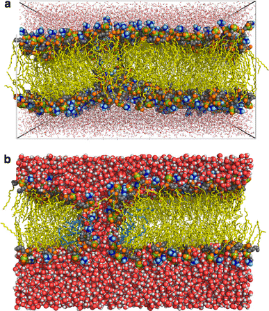
Pre-pore and pore in trajectories of a simulated bilayer under electrical tension. a Snapshot after 0.9 ns of MD simulation of a 1-palmitoyl-2-oleoylphosphatidylcholine (POPC) membrane (512 lipids) at electric field of 0.5 V/nm. Water is drawn as red sticks, lipid acyl chains are colored yellow, choline groups are in blue, phosphorus atoms are in green, lipid oxygens are in orange, and head-group carbons are in gray. Water-file defects are observed near the middle of the membrane. After 50 ns of simulation with a decreased field (V = 0.04 V/nm) the water wire has grown and transformed into a hydrophilic pore, shown in b (with the same color coding, and water in space-filling representation). The pore has an approximate radius of ~0.47 nm and is lined by ~8 lipids (highlighted light blue). Reprinted with permission from Biophysical Journal, Vol. 95 (Böckmann et al. 2008) © 2008 Biophysical Society
Fig. 5
Fig. 5
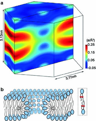
Bilayer structure at the edge of a peptide-induced lipid pore. The lipidic outline of the pore is viewed from the electron density of brominated lipids, resolved by grazing-angle X-ray diffraction (Qian et al. 2008b) a. The structure corresponds to the pore induced by the α5 active fragment of the protein Bax. It shows continuous electron density between the two monolayers at the pore edge, as expected for the case of toroidal pores with a wall lined (at least partially) by lipids, drawn schematically in b, where the bromine labels in the acyl chains of lipids are represented as small balls in lipids within a dashed-line box. Because these experiments do not provide data about the peptide part, we have purposely omitted it from the cartoon. A different pore-edge structure, with no participation of lipid head-groups in the wall, was found for the pore formed by alamethicin (a nonionic amphipathic peptide), which is in agreement with a barrel-stave model (Qian et al. 2008a). However, such a type of pore does not appear adequate for the majority of pore-active peptides, which are of cationic character, and it is thus not discussed in this work. The figure in A is reprinted with permission from PNAS, Volume 105 (Qian et al. 2008b) © 2008 National Academy of Sciences, USA
Fig. 6
Fig. 6
Free energy along the reaction coordinate for opening of a lipidic pore coupled to formation and reorganization of a peptide–membrane complex. Starting from peptide and membrane free species an initial membrane–peptide complex is formed favorably, with peptides adsorbed at the interface. As peptides accumulate, the membrane is stretched asymmetrically, causing thinning and increase of fluctuations. Peptides are postulated to bind more strongly in zones of bilayer defects, corresponding to the transition state, which reduces the activation free energy (∆G a) for lipid reorientation and lipidic pore formation. The ∆G a* energy barrier is expected to decrease with increasing the fraction of membrane-bound peptide up to some threshold value. Additionally, after the pore is formed, binding of the peptide near the pore rim stabilizes it via reduction of the line tension. Notice that the first part of the transformation resembles a catalyzed reaction
Fig. 7
Fig. 7
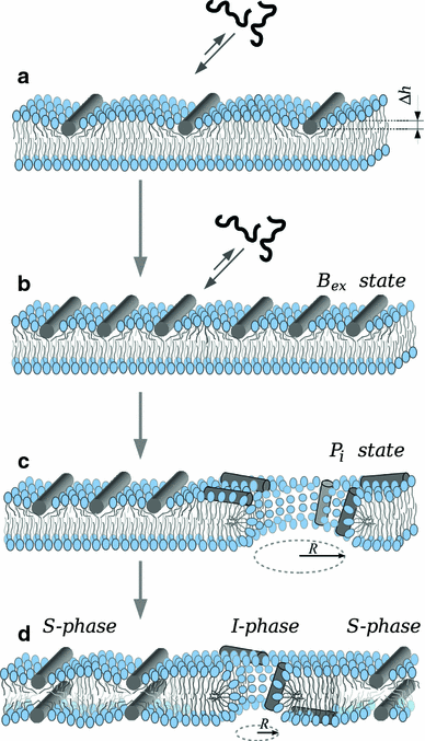
General mechanism for pore induction by cationic amphipathic peptides. a Pore-active peptides bind avidly to the accessible interface of lipid bilayers, even if they are made of zwitterionic lipids. Peptide binding is coupled to folding (in this case as an α-helix, represented by a cylinder). The peptides occupy a volume only in the interfacial region, causing asymmetric area stretching and membrane thinning (∆h). As a consequence, fluctuations increase and defects become more likely. b In the so-called B ex state (Tamba and Yamazaki 2009), membrane perturbation increases with the amount of bound peptides. We postulate (see the text and Fig. 6) that peptides bind with higher affinity near bilayer defects, stabilizing them and so reducing the activation energy for lipid reorganization and pore formation, which would constitute the transition state. c Eventually, the membrane yields and a pore is formed (so-called P i state by Tamba and Yamazaki). The initial pore is large (Tamba et al. ; Fuertes et al. 2010a). As the peptides diffuse through the pore to the opposite monolayer the accumulated asymmetric internal tension diminishes and the pore tends to close due to the line tension at the bilayer edge. However, the peptides bind near the pore rim and reduce the line tension, until an equilibrium is reached with a smaller but stable pore d. The appearance of pores can be understood as a phase transition, and in the equilibrium state two phases coexist (Huang et al. ; Huang 2009), namely S, with peptides bound essentially parallel to the membrane and lamellar bilayer lipids, and I, with peptides exhibiting a certain tilt rear the rim of pores and at the pore wall formed by lipid head groups. The structure of such a toroidal pore when induced by Bax-α5 has been characterized by grazing-angle X-ray diffraction (Qian et al. 2008b)
Fig. 8
Fig. 8
Shrinkage of pores induced by Bax-α5 on individual GUVs observed by fluorescence microscopy. a Bax-α5 and a first dye (Alexa555, blue) are initially together in the observation chamber, where GUVs (POPC:cardiolipin (CL) 80:20) labeled with 1,1-dioctadecyl-3,3,3,3-tetramethylindodicarbocyanine, 4-chlorobenzenesulfonate (DiD) (red) are added. A short time after (left panel), most observed GUVs are not yet porated. The leakage of the blue dye to the inside of GUVs is somewhat retarded and happens stochastically, but once initiated it completes for most GUVs in a few minutes (example kinetics are shown in b, graphs on the left). After 2 h, some GUVs are completely equilibrated with the outside dye while others are empty (a, middle panel). If then a second dye is added (Alexa 488, green), internalization occurs immediately and only for the vesicles completely porated at first instance (a, right panel). However, the entrance kinetics for this second leakage event is clearly slower (b, graphs in the right). In an independent experiment c the entry of Alexa555 (blue) and a fluorescein-labeled dextran of 10 kDa (FD10, green) induced by Bax-α5 on POPC:CL GUVs (80:20) were registered simultaneously by fluorescence microscopy. For any porated GUV, both dyes start leaking in at the same time and at a similar initial rate. However, the entrance of the larger FD10 is drastically slowed down before completion. Data from Fuertes et al. (2010a)

Similar articles

Cited by

References

    1. Abidor I, Arakelyan V, Chernomordik Y, Chizmadzhev V, Tarasevich M. Electrical breakdown ofBLM: main experimental facts and their qualitative discussion. Bioelectrochem Bioenerg. 1979;6:37–52.
    1. Ahmad S, Evans W. Post-translational integration and oligomerization of connexin 26 in plasma membranes and evidence of formation of membrane pores: implications for the assembly of gap junctions. Biochem J. 2002;365:693–699. - PMC - PubMed
    1. Almeida PF, Pokorny A. Mechanisms of Antimicrobial, Cytolytic, and Cell-Penetrating Peptides: from Kinetics to Thermodynamics. Biochemistry. 2009;48:8083–8093. - PMC - PubMed
    1. Andersen S, Jackson A, Heimburg T. Towards a thermodynamic theory of nerve pulse propagation. Prog Neurobiol. 2009;88:104–113. - PubMed
    1. Antonov VF, Petrov VV, Molnar AA, Predvoditelev DA, Ivanov AS. The appearance of single-ion channels in unmodified lipid bilayer membranes at the phase transition temperature. Nature. 1980;283:585–586. - PubMed

Publication types

MeSH terms

LinkOut - more resources