. 2011 Mar;49(3):124-33.
doi: 10.1002/dvg.20695. Epub 2011 Jan 5.

Afp::mCherry, a red fluorescent transgenic reporter of the mouse visceral endoderm

Affiliations

Afp::mCherry, a red fluorescent transgenic reporter of the mouse visceral endoderm

Manuel Viotti et al. Genesis. 2011 Mar.

Abstract

Live imaging of genetically encoded fluorescent protein reporters is increasingly being used to investigate details of the cellular behaviors that underlie the large-scale tissue rearrangements that shape the embryo. However, the majority of mouse fluorescent reporter strains are based on the green fluorescent protein (GFP). Mouse reporter strains expressing fluorescent colors other than GFP are therefore valuable for co-visualization studies with GFP, where relative positioning and relationship between two different tissues or compartments within cells are being investigated. Here, we report the generation and characterization of a transgenic Afp::mCherry mouse strain in which cis-regulatory elements from the Alpha-fetoprotein (Afp) locus were used to drive expression of the monomeric Cherry red fluorescent protein. The Afp::mCherry transgene is based on and recapitulates reporter expression of a previously described Afp::GFP strain. However, we note that perdurance of mCherry protein is not as prolonged as GFP, making the Afp::mCherry line a more faithful reporter of endogenous Afp expression. Afp::mCherry transgenic mice expressed mCherry specifically in the visceral endoderm and its derivatives, including the visceral yolk sac, gut endoderm, fetal liver, and pancreas of the embryo. The Afp::mCherry reporter was also noted to be expressed in other documented sites of Afp expression including hepatocytes as well as in pancreas, digestive tract, and brain of postnatal mice.


Figures

FIG. 1
FIG. 1. Design and validation of the Afp::mCherry transgene in cells
(a) Schematic representation of the Afp::mCherry construct. (b) Transfection of the pAfp::mCherry plasmid in cells expressing Afp endogenously resulted in expression of the mCherry reporter. Upper row depicts low magnification views of fluorescence overlayed onto the brightfield channel, lower row depicts high magnifications views of fluorescent channel. Hepa1-6, HepG2, and Caco-2 cells transfected with the plasmids pAfp::mCherry and Afp::GFP contained both red and green fluorescent cells. Cells were counterstained with Hoechst (blue). Scale bars represent 20 μm.
FIG. 2
FIG. 2. Afp::mCherry transgene activity is detected throughout the visceral endoderm and its derivatives
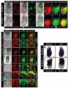
(a) In PS stage (E6.0) Afp::mCherrytg/+; Afp::GFPtg/+ embryos, mCherry co-localized with GFP and was expressed throughout the VE with somewhat heterogenous intensities (both in emVE and exVE, except the most proximal part adjacent to the ectoplacental cone, where both fluorescent proteins are absent). (b) The overall correlation of mCherry and GFP expression was maintained at the ES (E6.5) stage. (c) Lateral view of an EB (E7.25) Afp::mCherrytg/+; Afp::GFPtg/+ embryo depicting mCherry-positive emVE cells and uniform fluorescence in the exVE, with identical GFP localization. (c′) High magnification of early dispersal stage of VE, showing faithful co-expression of red and green fluorescence. (d) Anterior view depicting co-localization along the anterior midline. (e) Lateral view of LB (E7.75) stage Afp::mCherrytg/+; Afp::GFPtg/+ embryo showing red fluorescence in the exVE and dispersed emVE. (e′) High magnification view of late dispersal stage of VE indicating co-localization of red and green fluorescence in the majority of cells (white arrowheads) and a subset of GFP-positive cells with low levels of mCherry (orange arrowheads). (f,g) Anterior and posterior views of the distal section of embryo overlying the epiblast indicating weaker or absence of red fluorescence in a subset of GFP-positive cells (orange arrowheads). (h) Whole-mount in situ hybridization in ES stage (E6.5) Afp::mCherrytg/+ embryos revealed mCherry transcript present throughout VE (both emVE and exVE), in agreement with the localization of GFP transcripts in stage-matched Afp::GFPtg/+ embryos. (i) By the LB stage (E7.5) mCherry transcripts appeared restricted to the exVE in Afp::mCherrytg/+ embryos, again corresponding to the expression pattern of GFP in Afp::GFP tg/+ embryos. Pr, proximal; D, distal; A, anterior; P, posterior; 2D, 2-dimensions; 3D, 3-dimensions. Scale bars represent 20 μm in a and b and 50 μm in c-i.
FIG. 3
FIG. 3. Organization of mCherry-positive cells around the node and notochordal plate
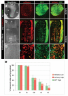
(a) Lateral view of an Afp::mCherrytg/+; Afp::GFPtg/+ embryo at the 5-6 somite stage (E8.5) showing widespread red fluorescence in the visceral yolk sac. (b) Ventral view revealing stereotypical organization of mCherry-positive cells around the node and bilaterally along the notochordal plate. (b′) High magnification view indicating absence (blue arrowheads) or low levels (orange arrowheads) of mCherry in a subset (~50%) (percentage adjusted after quantitation) of GFP-positive cells. (c) Histogram (mean ± sd) depicting percent fluorescent cells overlying the epiblast. Fluorescence was measured using Volocity software, where ‘high’ fluorescence was determined as above fluorescent intensity of 50, and ‘low’ fluorescence was considered in the range between intensity of 50 and background levels. N=3 embryos for each stage. Pr, proximal; D, distal; A, anterior; P, posterior; fg, foregut invagination; som, somites; hg, hindgut invagination. Scale bars represent 100 μm.
FIG. 4
FIG. 4. Afp::mCherry expression in the ~15 somite stage embryo
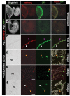
(a) Lateral wholemount view of Afp::mCherrytg/+; Afp::GFPtg/+ E9.0 embryo illustrating widespread red and green fluorescence in the yolk sac. (b) View of the same embryo but in isolation from the yolk sac, allowing detection of green and red fluorescence along the gut tube. (c) Section through the yolk sac indicating localization of mCherry specifically in the endoderm layer. Cells of the endoderm were mostly positive for red and green fluorescence (white arrowheads) while yolk sac mesoderm cells of the yolk sac were non-fluorescent (purple arrowheads). (d-e) Sections through foregut and midgut regions revealed cells in the endodermal lining of the gut tube lumen being positive for GFP with low levels of mCherry (orange arrowheads). (f) In hindgut sections some endoderm cells were positive for both fluorescent proteins (white arrowheads), while other cells were GFP-positive, with low or undetectable levels of mCherry (orange arrowheads). fg, foregut invagination; som, somites; hg, hindgut invagination. Scale bars represent 50 μm.
FIG. 5
FIG. 5. Additional sites of mCherry localization during organogenesis
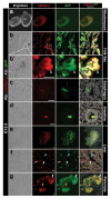
(a) Lateral wholemount view of an Afp::mCherrytg/+; Afp::GFPtg/+ embryo at E12.5 indicating red and green fluorescent reporter expression in the yolk sac. (b-g) Sections taken through the embryo in (a) revealing Afp::mCherry transgene activity in endoderm derivatives. (b) Section through the yolk sac revealed that the majority of GFP-positive cells were also mCherry-positive. (b′) High magnification of the section of yolk sac indicating that fluorescence is specific to the endoderm layer. Fluorescent cells (white arrowheads) form a contiguous epithelium distinct from a second, non-fluorescent layer (purple arrowheads). (c,d) Sections of gut tube derivatives with double fluorescent cells. (e,f) The liver of Afp::mCherrytg/+; Afp::GFPtg/+ embryos at this stage expressed both mCherry and GFP, yet these reporters did not necessarily co-localize at a cellular level. Some liver cells were non-fluorescent, some were only mCherry-positive (pink arrowheads), some were only GFP-positive (blue arrowhead) while others were positive for both reporters (white arrowheads). (g) Section of the pancreatic bud reveals that all cells were GFP-positive, a large number were also mCherry-positive (white arrowheads) and in a subset of cells mCherry was either present at low levels or absent (orange arrowheads). Scale bars represent 100 μm.
FIG. 6
FIG. 6. Localization of mCherry in organs of newborn mice
(a) Section through an Afp::mCherrytg/+; Afp::GFPtg/+ P3 brain revealing cells positive for both fluorescent protein reporters. (b) Wholemount view of the newborn stomach revealing red fluorescence in GFP-positive cells. (c) Wholemount image of the liver indicating widespread fluorescence of both GFP and mCherry. (c′-c″) Sections through the liver depicted in (c) revealed extensive, but not always correlative reporter expression. (d) Wholemount view of the newborn pancreas showing broad coincidence of GFP and mCherry. (d′) Section through the pancreas indicating mCherry fluorescence in the majority of GFP-positive cells. (e) Wholemount view of the newborn intestine indicating extensive regions of reporter co-localization. (e′-e″) Sections through the intestine revealing cells along the villi positive for both GFP and mCherry (white arrowheads), or GFP-positive with low or undetectable levels of mCherry (orange arrowheads). Scale bars represent 500 μm in c′ and e′, and 100 μm in all other panels.

References

    1. Campbell RE, Tour O, Palmer AE, Steinbach PA, Baird GS, Zacharias DA, Tsien RY. A monomeric red fluorescent protein. Proc Natl Acad Sci USA. 2002;99:7877–7882. - PMC - PubMed
    1. Downs KM, Davies T. Staging of gastrulating mouse embryos by morphological landmarks in the dissecting microscope. Development. 1993;118:1255–1266. - PubMed
    1. Dziadek M. Modulation of alphafetoprotein synthesis in the early postimplantation mouse embryo. J Embryol Exp Morphol. 1978;46:135–146. - PubMed
    1. Dziadek M, Adamson E. Localization and synthesis of alphafoetoprotein in post-implantation mouse embryos. J Embryol Exp Morphol. 1978;43:289–313. - PubMed
    1. Ferrer-Vaquer A, Viotti M, Hadjantonakis AK. Transitions between epithelial and mesenchymal states and the morphogenesis of the early mouse embryo. Cell Adh Migr. 2010;4:447–457. - PMC - PubMed

Publication types

MeSH terms