. 2011 Apr 28;54(8):2714-26.
doi: 10.1021/jm101505d. Epub 2011 Mar 28.

A potent and orally active antagonist (SM-406/AT-406) of multiple inhibitor of apoptosis proteins (IAPs) in clinical development for cancer treatment

Affiliations

A potent and orally active antagonist (SM-406/AT-406) of multiple inhibitor of apoptosis proteins (IAPs) in clinical development for cancer treatment

Qian Cai et al. J Med Chem. .

Abstract

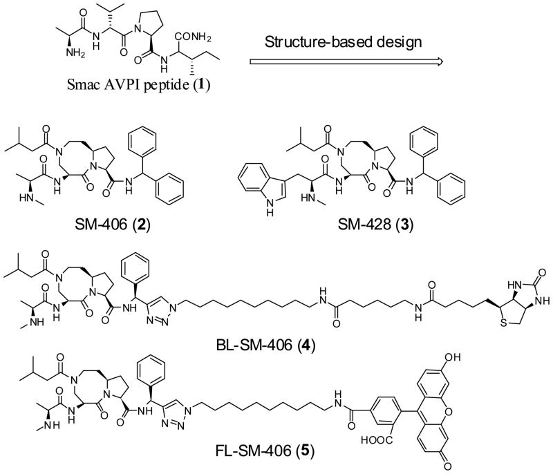
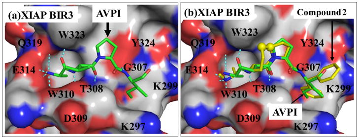
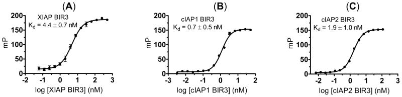
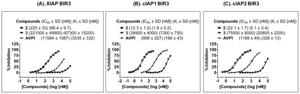
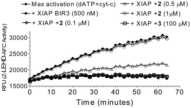
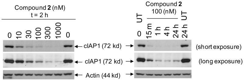
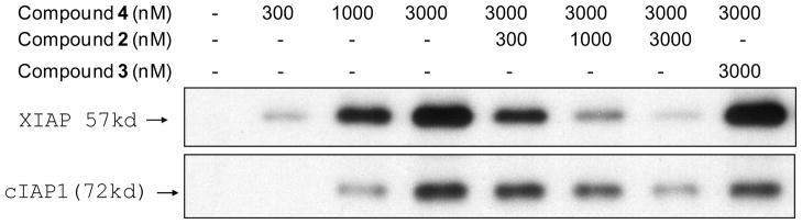
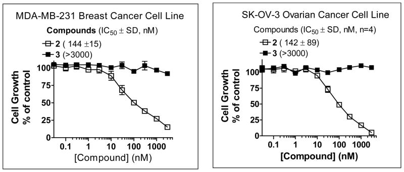
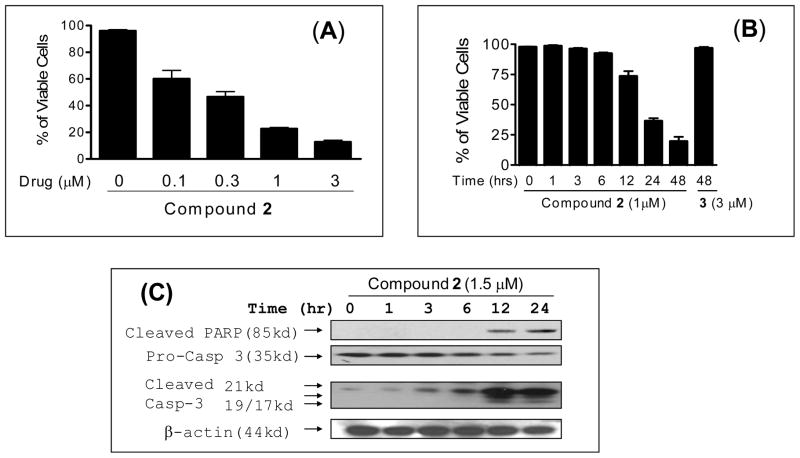
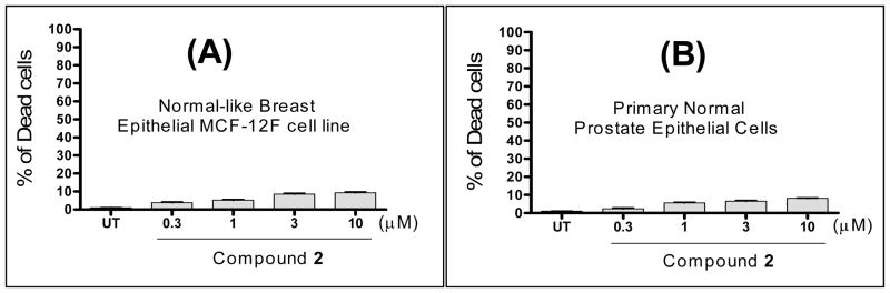
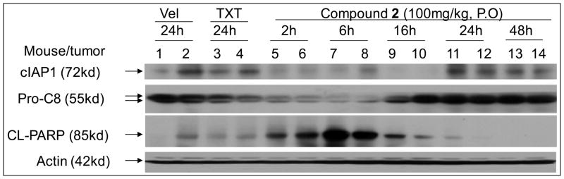
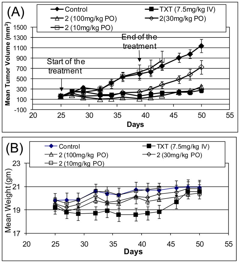
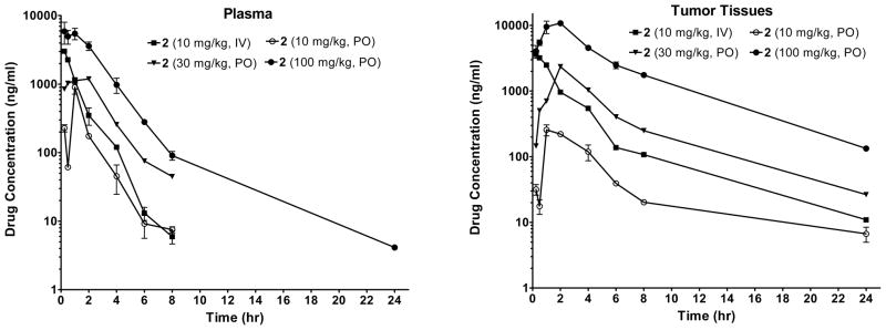
We report the discovery and characterization of SM-406 (compound 2), a potent and orally bioavailable Smac mimetic and an antagonist of the inhibitor of apoptosis proteins (IAPs). This compound binds to XIAP, cIAP1, and cIAP2 proteins with K(i) of 66.4, 1.9, and 5.1 nM, respectively. Compound 2 effectively antagonizes XIAP BIR3 protein in a cell-free functional assay, induces rapid degradation of cellular cIAP1 protein, and inhibits cancer cell growth in various human cancer cell lines. It has good oral bioavailability in mice, rats, non-human primates, and dogs, is highly effective in induction of apoptosis in xenograft tumors, and is capable of complete inhibition of tumor growth. Compound 2 is currently in phase I clinical trials for the treatment of human cancer.


Figures

Figure 1
Figure 1
Chemical structures of Smac AVPI peptide, a potent and orally active Smac mimetic 2, its inactive control 3, a biotinylated analogue 4 and a fluorescently tagged analogue 5.
Figure 2
Figure 2
Structure-based design of conformationally constrained Smac mimetics. (a). Crystal structure of Smac in a complex with XIAP BIR3. For clarity, only the AVPI peptide in Smac is shown. (b). Superposition of the predicted binding model of compound 2 in a complex with BIR3 on the crystal structure of Smac AVPI peptide complexed with XIAP BIR3. The carbon atoms of AVPI peptide and compound 2 are shown in yellow and green, respectively; the nitrogen and oxygen atoms in both ligands are shown in blue and red, respectively. The protein is represented as its solvent accessible surface and colored by atom types (carbon: gray; nitrogen: blue; oxygen: red). Hydrogen bonds are shown in dashed light blue lines.
Figure 3
Figure 3
Saturation curves of fluorescently tagged compound 5 to (A). XIAP BIR3 protein; (B). cIAP1 BIR3 protein; and (C). cIAP2 BIR3 protein. The standard deviation for the Kd value to each protein was calculated from three independent experiments.
Figure 4
Figure 4
Determination of the binding affinities of compounds 2, 3 and the Smac AVPI peptide to (A). XIAP BIR3; (B). cIAP1 BIR3; and (C). cIAP2 BIR3 using sensitive and quantitative fluorescence-polarization assays. The standard deviation for the IC50 value to each protein was calculated from three independent experiments.
Figure 5
Figure 5
Functional antagonism of compound 2 against XIAP BIR3 to restore caspase-9 activity in a cell-free assay. Compound 3 was used as the control compound.
Figure 6
Figure 6
Compound 2 induces rapid cIAP-1 degradation in the MDA-MB-231 cancer cell line. Cells were treated with compound 2 as indicated and Western blot analysis was performed using specific antibody against cIAP1 and Actin.
Figure 7
Figure 7
Biotin-streptavidin pull-down experiments to probe the binding of compounds 2 and 3 to cellular XIAP and cIAP1 in the MDA-MB-231 breast cancer cell lysates using biotinylated compound 4.
Figure 8
Figure 8
Inhibition of cell growth by compounds 2 and 3 in the MDA-MB-231 breast cancer and SK-OV-3 ovarian cancer cell lines, as determined in a water soluble tetrazolium (WST) cell proliferation assay. Four independent experiments were performed for each compound in each cell line.
Figure 9
Figure 9
Analysis of induction of cell death and apoptosis by compound 2 in the MDA-MB-231 cell line. MDA-MB-231 cells were treated with compound 2 at (A) different concentrations for 24 hours (B) with 1 μM concentration for different time points. (C) Western blot analysis of caspase processing and cleavage of poly (ADP-ribose) polymerase (PARP). MDA-MB-231 cells were treated with 1.5 μM concentration of compound 2 for 24 hours and cleaved PARP, pro-caspase-3 and cleaved caspase-3 were probed with specific antibodies and actin was used as a loading control.
Figure 10
Figure 10
Induction of cell death by compound 2 in (A): normal-like human breast epithelial MCF-12F cells. (B): Primary human normal prostate epithelial cells; Cells were treated with different concentrations of compound 2 for 48 hours and cell viability was determined using the trypan blue exclusion assay.
Figure 11
Figure 11
Evaluation of compound 2 on cIAP1, caspase-8 processing and PARP cleavage in the MDA-MB-231 xenograft tissues. Mice bearing the MDA-MB-231 tumors were treated with a single oral dose of compound 2, intravenous taxotere (TXT) or intravenous vehicle. cIAP1, XIAP, pro-caspase-8 (Pro-C8) and cleaved PARP (CL-PARP) were examined by Western blotting.
Figure 12
Figure 12
In vivo antitumor activity of compound 2 in the MDA-MB-231 xenograft model. Taxotere (TXT) was used as the control. (A). Tumor volume. (B). Animal weight.
Figure 13
Figure 13
Analysis of pharmacokinetics of compound 2 in plasma and in tumor tissues in SCID mice bearing the MDA-MB-231 xenograft tumors. Compound 2 was dosed via either intravenously (IV) or oral gavage (PO).
Scheme 1
Scheme 1
Synthesis of compounds 2–5 Reagents and conditions: (a) i. isovaleryl chloride, N,N-diisopropylethylamine, CH2Cl2; ii. 2 N LiOH, 1,4-dioxane, 82% over two steps; (b) aminodiphenylmethane, EDC, HOBt, N,N-diisopropylethylamine, CH2Cl2, 83%; (c) i. 4 N HCl in 1,4-dioxane, methanol; ii. L-N-Boc-N-methyl-alanine, EDC, HOBt, N,N-diisopropylethylamine, CH2Cl2; iii. 4 N HCl in 1,4-dioxane, methanol, 74% over three steps; (d) i. 4 N HCl in 1,4-dioxane, methanol; ii. L-Boc-Na-methyl-tryptophan, EDC, HOBt, N,N-diisopropylethylamine, CH2Cl2; iii. 4 N HCl in 1,4-dioxane, methanol, 63% over three steps; (e) i. Z-6-aminohexanoic acid, EDC, HOBt, N,N-diisopropylethylamine, CH2Cl2; ii. MsCl, Et3N, CH2Cl2; iii. NaN3, DMF, 110°C, 49% over three steps; (f) i. S-1-phenylprop-2-yn-1-amine, EDC, HOBt, N,N-diisopropylethylamine, CH2Cl2; ii. 4 N HCl in 1,4-dioxane, methanol; iii. L-N-Boc-N-methyl-alanine, EDC, HOBt, N,N-diisopropylethylamine, 69% over three steps; (g) i. 10, CuSO4, sodium L-ascorbate, t-BuOH-H2O 1:1; ii. H2, 10% Pd-C, methanol; iii. (+)-biotin N-hydroxy-succinimide ester, N,N-diisopropylethylamine, CH2Cl2; iv. 4 N HCl in 1,4-dioxane, MeOH, 43% over four steps; (h) i. CbzCl, Et3N, CH2Cl2; ii. MsCl, Et3N, CH2Cl2; iii. NaN3, DMF, 66% over three steps; i. 11, CuSO4, sodium L-ascorbate, t-BuOH-H2O 1:1; ii. H2, 10% Pd-C, methanol; iii. 5-Carboxyfluorescein N-succinimidyl ester, N,N-diisopropylethylamine, CH2Cl2; iv. 4 N HCl in 1,4-dioxane, MeOH, 34% over four steps.

Similar articles

Cited by

References

    1. Nicholson DW. From bench to clinic with apoptosis-based therapeutic agents. Nature. 2000;407:810–816. - PubMed
    1. Ponder BA. Cancer genetics. Nature. 2001;411:336–341. - PubMed
    1. Lowe SW, Lin AW. Apoptosis in cancer. Carcinogenesis. 2000;21:485–495. - PubMed
    1. Hanahan D, Weinberg RA. The hallmarks of cancer. Cell. 2000;100:57–70. - PubMed
    1. Salvesen GS, Duckett CS. Apoptosis: IAP proteins: blocking the road to death’s door. Nat Rev Mol Cell Biol. 2002;3:401–410. - PubMed

Publication types

MeSH terms

Substances

LinkOut - more resources