Review
. 2011 Jun;23(6):542-53.
doi: 10.1111/j.1365-2826.2011.02130.x.

Physiology of the gonadotrophin-releasing hormone (GnRH) neurone: studies from embryonic GnRH neurones

Affiliations
Review

Physiology of the gonadotrophin-releasing hormone (GnRH) neurone: studies from embryonic GnRH neurones

S Constantin. J Neuroendocrinol. 2011 Jun.

Abstract

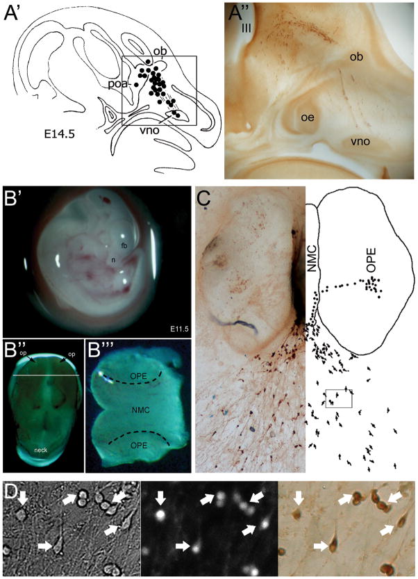
Gonadotrophin-releasing hormone (GnRH)-secreting neurones are the final output of the central nervous system driving fertility in all mammals. Although it has been known for decades that the efficiency of communication between the hypothalamus and the pituitary depends on the pulsatile profile of GnRH secretion, how GnRH neuronal activity is patterned to generate pulses at the median eminence is unknown. To date, the scattered distribution of the GnRH cell bodies remains the main limitation to assessing the cellular events that could lead to pulsatile GnRH secretion. Taking advantage of the unique developmental feature of GnRH neurones, the nasal explant model allows primary GnRH neurones to be maintained within a micro-network where pulsatile secretion is preserved and where individual cellular activity can be monitored simultaneously across the cell population. This review summarises the data obtained from work using this in vitro model, and brings some insights into GnRH cellular physiology.


Figures

Figure 1
Figure 1. GnRH neuron migration is maintained in nasal explants, allowing cellular physiological studies on native GnRH neurons
A′. Diagram showing the migratory track of GnRH neurons in a E14.5 mouse, from the vomeronasal organ (vno) to the rostral preoptic area (poa), entering the brain with the olfactory fibers at the level of the olfactory bulbs (ob) (49); A″. Photomicrograph boxed area in A Section has been immunocytochemically stained for GnRH (oe, olfactory epithelium). B. Preparation of nasal explants at E11.5 (B′, n: nose, fb: forebrain); B″, Isolated head sitting on its top, displaying the nasal placodes (op); B‴, The tip of the nose sectioned where the nasal midline cartilage (NMC) and the olfactory placode epithelium (OPE) are delimited. C. Representative picture of a nasal explant after 7 days in culture and the mirror corresponding diagram showing the differentiated GnRH-expressing cells in the OPE, migrating towards the NMC and emerging outside the tissue mass as they migration. D. Representative picture of GnRH neurons used for calcium imaging in an area corresponding to the box delimited in (C). Left panel, bright field; Middle panel, calcium dye-loaded cells; Right panel, post hoc identification by immunocytochemistry for GnRH. Arrows indicate the identical cells over the procedure. [Adapted with permission from (49) for A & (43) for A′, (63) for D; pictures for B were generously provided by Dr. P. Giacobini].
Figure 2
Figure 2. GnRH cells in nasal explant display spontaneous rhythmic activity
A. Example of a secretion profile illustrating in vitro release of GnRH with pulsatile pattern (~ 1 pulse/20min). B. Simultaneous recording of electrical activity and intracellular calcium level in a single GnRH neuron revealed spontaneous bursts of activity time-correlated with transient oscillations in [Ca2+]i. C. Simultaneous recordings of a cohort of GnRH neurons illustrating the occurrence of [Ca2+]i oscillations (white ticks) being independent in each cell (one line/cell) (upper panel) but synchronized across the GnRH cells every ~ 20min (lower panel). [Adapted with permission from (55) for A, (63) for B–C].
Figure 3
Figure 3. Three different downstream pathways of G-protein coupled receptors can modulate GnRH neuronal activity
A. Forskolin-evoked increase in intracellular cAMP, modeling the activation of Gs pathway, stimulates GnRH neuronal activity. B. Stimulation can be directly mimicked with Sp-cAMPS, a selective activator of protein kinase A. C–D. Neuropeptide Y inhibits GnRH neuronal activity. TPN-Q prevents the NPY-evoked inhibition, indicating a functional Gi pathway coupled to G-inward rectifier potassium channels (GIRK). E. Kisspeptin-10 induces a potent stimulation. F. A similar response can be induced directly with the phorbol ester PMA, an activator of PKC. [Adapted with permission from - (64) for A–B, (99) for C–D, (65) for E–F]. [TTX=tetrodotoxin 1 μM, voltage-gated sodium channel blocker; BIC=bicuculline 20 μM, A-subtype GABAergic receptor antagonist; CNQX=6-cyano-7-nitroquinoxaline-2,3-dione 10 μM, AMPA-subtype glutamatergic receptor antagonist; PRG=proglumide 100 nM, cholecystokininergic receptor antagonist].
Figure 4
Figure 4. The rhythmic activity of GnRH cells in nasal explants arises from different cell populations
A. Recording of [Ca2+]i in a single GnRH neuron illustrating spontaneous oscillations can persist in presence of TTX. The [Ca2+]i oscillations are partially blocked by bicuculline (B), CNQX (C) and proglumide (D) revealing GABAergic, glutamatergic and cholecystokininergic inputs, respectively. Together, constitutively active endogenous inputs are responsible for the rhythmic activity displayed by GnRH neurons (E). [Adapted with permission from (68)]. [FSK=forskolin 1 μM, adenylyl cyclase activator; Sp-cAMPS=Sp-adenosine-3,5-cyclic monophosphorothioate triethylammonium 100 nM, protein kinase A activator; NPY=neuropeptide-Y 1 nM, NPY receptor agonist; TPN-Q=tertiapin-Q 100 nM, Gi-activated inward rectifier potassium channel blocker; KP-10=kisspeptin-10 10 nM, GPR54 receptor agonist; BIC=bicuculline 20 μM, A-subtype GABAergic receptor antagonist; PMA=phorbol 12-myristate 13-acetate 50 nM, protein kinase C activator].
Figure 5
Figure 5. Schematic of the endogenous inputs modulating GnRH neurons and their signaling pathways
Action potentials induce the activity of the calcium oscillator, resulting in a transient increase in [Ca2+]i via voltage-gated calcium channels and yet to be identified calcium source. GABAergic, glutamatergic and cholecystokininergic inputs, the major inputs present in vitro, are able to trigger the calcium oscillator via the activation of GABAA, AMPA and CCK1 receptors, respectively. Gi-coupled receptors can reduce the activity of the oscillator via a direct coupling to GIRK channels while Gs-coupled receptors can stimulate GnRH neurons by increasing the activity of protein kinase A upon the calcium oscillator. Gq-coupled receptors can stimulate GnRH neuronal calcium oscillator by at least two mechanisms initiated by the activity of phospholipase C: protein kinase C and transient receptor potential channels.

References

    1. King JC, Tobet SA, Snavely FL, Arimura AA. LHRH immunopositive cells and their projections to the median eminence and organum vasculosum of the lamina terminalis. J Comp Neurol. 1982;209(3):287–300. - PubMed
    1. Hoffman GE, Phelps CJ, Khachaturian H, Sladek JR., Jr . Neuroendocrine Projections to the Median Eminence. In: Ganten D, Pfaff D, editors. Morphology of Hypothalamus and Its Connections. Berlin - Heidelberg - New York - London -Paris - Tokyo: Academic Press; 1986. pp. 161–96.
    1. Silverman AJ, Jhamandas J, Renaud LP. Localization of luteinizing hormone-releasing hormone (LHRH) neurons that project to the median eminence. J Neurosci. 1987;7(8):2312–9. - PMC - PubMed
    1. McCann SM, Ramirez VD. The Neuroendocrine Regulation of Hypophyseal Luteinizing Hormone Secretion. Recent Prog Horm Res. 1964:20131–81. - PubMed
    1. Spergel DJ, Kruth U, Shimshek DR, Sprengel R, Seeburg PH. Using reporter genes to label selected neuronal populations in transgenic mice for gene promoter, anatomical, and physiological studies. Prog Neurobiol. 2001;63(6):673–86. - PubMed

Publication types

Substances