. 2011 Mar 28;13(2):R54.
doi: 10.1186/ar3298.

Cystatin C influences the autoimmune but not inflammatory response to cartilage type II collagen leading to chronic arthritis development

Affiliations

Cystatin C influences the autoimmune but not inflammatory response to cartilage type II collagen leading to chronic arthritis development

Alexandra Bäcklund et al. Arthritis Res Ther. .

Abstract

Introduction: Collagen-induced arthritis (CIA) is a mouse model for rheumatoid arthritis (RA) and is induced after immunization with type II collagen (CII). CIA, like RA, is an autoimmune disease leading to destruction of cartilage and joints, and both the priming and inflammatory phases have been suggested to be dependent on proteases. In particular, the cysteine proteases have been proposed to be detrimental to the arthritic process and even immunomodulatory. A natural inhibitor of cysteine proteases is cystatin C.

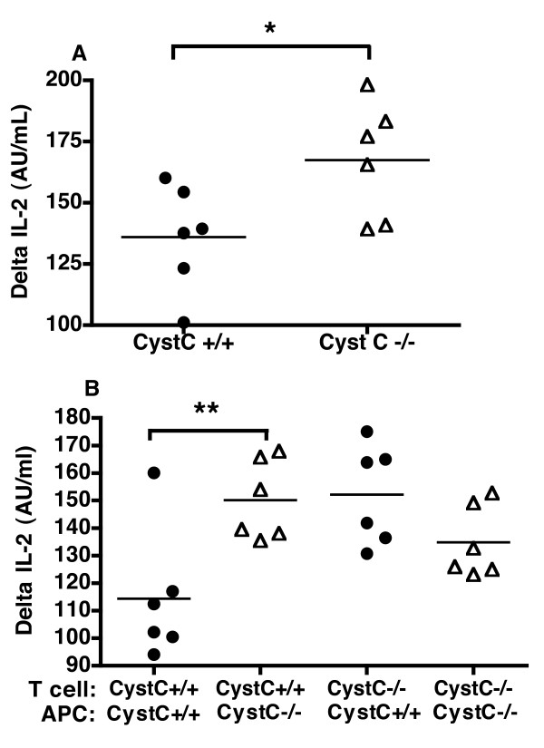
Methods: Cystatin C-deficient, sufficient and heterozygous mice were tested for onset, incidence and severity of CIA. The effect of cystatin C-deficiency was further dissected by testing the inflammatory effector phase of CIA; that is, collagen antibody-induced arthritis model and priming phase, that is, T cell response both in vivo and in vitro. In addition, in order to determine the importance of T cells and antigen-presenting cells (APCs), these cell populations were separated and in vitro T cell responses determined in a mixed co-culture system. Finally, flow cytometry was used in order to further characterize cell populations in cystatin C-deficient mice.

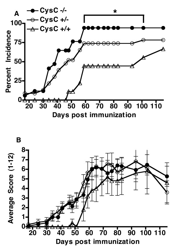
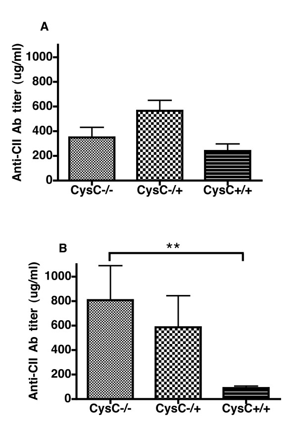
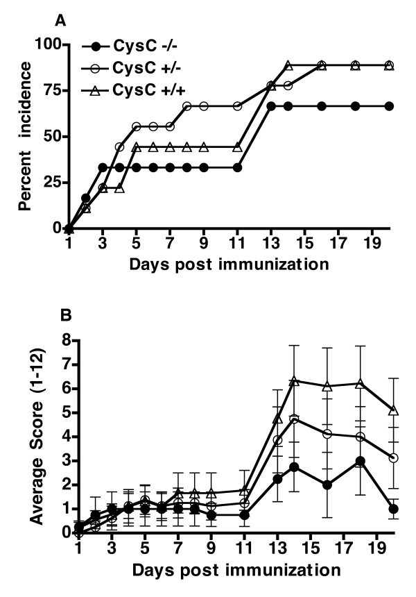
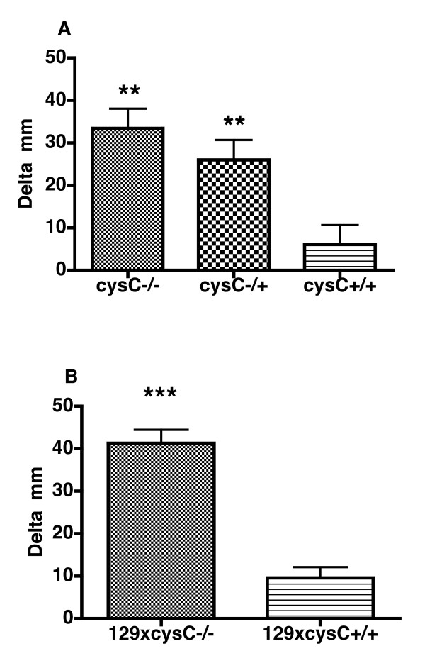
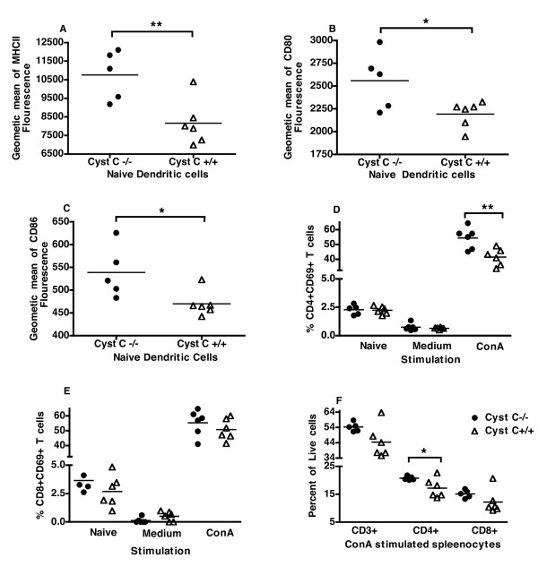
Results: Here, we show that mice lacking cystatin C, develop arthritis at a higher incidence and an earlier onset than wild-type controls. Interestingly, when the inflammatory phase of CIA was examined independently from immune priming then cystatin C-deficiency did not enhance the arthritis profile. However, in line with the enhanced CIA, there was an increased T cell and B cell response as delayed-type hypersensitivity reaction and anti-CII antibody titers were elevated in the cystatin C-deficient mice after immunization. In addition, the ex vivo naïve APCs from cystatin C-deficient mice had a greater capacity to stimulate T cells. Interestingly, dendritic cells had a more activated phenotype in naïve cystatin C-deficient mice.

Conclusions: The lack of cystatin C enhances CIA and primarily affects in vivo priming of the immune system. Although the mechanism of this is still unknown, we show evidence for a more activated APC compartment, which would elevate the autoimmune response towards CII, thus resulting in an enhanced development of chronic arthritis.


Figures

Figure 1
Figure 1
Cystatin C-deficient mice have a higher incidence of arthritis but have a similar arthritic score. A, Cystatin C-deficient mice (Cyst C-/-, n = 17) had a greater cumulated arthritis incidence (that is, all mice that have shown signs of arthritis at any time point are included as diseased individuals) compared to heterozygous (Cyst C-/+, n = 23) and wild type litters Cyst C+/+, n = 9). Data shown are from one representative experiment out of two separate experiments (combined data shown in Table 1). * P-value < 0.05 as tested in Fisher's exact test. B, Cystatin C-deficient mice (n = 15) had a similar arthritic severity compared to heterozygous (n = 17) and wild type littermate controls (n = 5) when affected mice were compared (mice were excluded if no clinical signs of arthritis was seen after 115 days post immunization).
Figure 2
Figure 2
The anti-CII IgG antibody titers are elevated in the cystatin C-deficient mice. Mice were bled at Day 34 (A) before boost immunization with CII/IFA, and at the end of the experiment at Day 115 (B). There was a statistically significant difference between the antibody production of cystatin C-deficient mice (Cyst C -/-) and the wild type controls (Cyst C +/+) at Day 115. ** P-value < 0.01 as tested with Mann-Whitney.
Figure 3
Figure 3
Effector phase is similar between cystatin C-deficient and control mice. CAIA was induced with two monoclonal anti-CII antibodies by i.v injection into cystatin C-deficient (Cyst C -/-, n = 6), cystatin C heterozygous (Cyst C -/+, n = 9) and WT littermate controls (Cyst C +/+, n = 10). A, No significant difference was seen in incidence of arthritis. B, There was also no difference in arthritis severity when comparing affected mice (Cyst C -/- n = 4, Cyst C -/+ n = 8, Cyst C +/+ n = 9, mice were excluded if no clinical signs of arthritis was seen after 20 days post antibody injection). The error bars indicate SEM.
Figure 4
Figure 4
Cystatin C-deficient mice show a stronger DTH reaction. A, Cystatin C-deficient mice (Cyst C -/-, n = 11) developed a significantly stronger DTH reaction towards CII compared to the wild type controls (Cyst C +/+, n = 11) 48 h after challenge, and so did the heterozygous cystatin C-deficient mice (Cyst C -/+, n = 11). B, The observed enhanced DTH was due to cystatin C-deficiency and not 129/Sv linked genes as F1 intercross between 129/Sv x cystatin C-deficient mice (n = 12) had a significantly enhanced DTH compared to the F1 intercross between 129/Sv x B10.Q/rhd (n = 12) 48 h after challenge. Significance was calculated with Mann-Whitney test where ** P-value < 0.01 and *** P-value < 0.001.
Figure 5
Figure 5
Cystatin C-deficient APCs have a greater propensity to stimulate cystatin C-sufficient T cells. Splenocytes were prepared from either cystatin C-deficient mice or wild type mice and stimulated in vitro with either media alone or with 2 ug/mL ConA (change in production level (delta) is shown, where the response to media alone is deducted from the ConA response). Cystatin C-deficient splenocytes mice (CysC-/-) had a significantly higher production of IL-2 (A) after 24 h Cystatin C-deficient APCs were able to enhance the IL-2 production of T cells from wild type mice, where as there was no difference in IL-2 production in cystatin C-deficient T cells when co-cultured with either wild type (CysC+/+) or cystatin C-deficient APCs (B). However, when wild type APCs where co-cultured with T cells then the cystatin C-deficient T cells produced more IL-2 than wild type T cells. Six mice were used per group, statistical significance was calculated by Students t-test where * P-value < 0.05 and ** P-value < 0.01.
Figure 6
Figure 6
Cystatin C-deficient DCs have increased expression of MHCII and Co-stimulatory molecules, and more activated T cells. Splenocytes from naïve mice or splenocytes stimulated with ConA or media were analyzed by flow cytometry. Naive DCs from cystatin C-deficient (CysC-/-) mice expressed higher intensity of MHCII (A) CD80 (B) and CD86 (C). A greater percent of CD4+ T cells expressed CD69 in cystatin C-deficient mice than wild type mice (CysC+/+) when stimulated with ConA for 24 h (D) but not CD8+ T cells. (E), In line with this there was a higher percent of CD4+ T cells in cystatin C-deficient mice and a tendency for a higher percent of CD8+ and total T cells (F). Six mice were used per group, statistical significance was calculated by Students t-test where * P-value < 0.05 and ** P-value < 0.01.

References

    1. Gabrijelcic D, Annan-Prah A, Rodic B, Rozman B, Cotic V, Turk V. Determination of cathepsins B and H in sera and synovial fluids of patients with different joint diseases. J Clin Chem Clin Biochem. 1990;28:149–153. - PubMed
    1. Lenarcic B, Gabrijelcic D, Rozman B, Drobnic-Kosorok M, Turk V. Human cathepsin B and cysteine proteinase inhibitors (CPIs) in inflammatory and metabolic joint diseases. Biol Chem Hoppe Seyler. 1988;369(Suppl):257–261. - PubMed
    1. Trabandt A, Gay RE, Fassbender HG, Gay S. Cathepsin B in synovial cells at the site of joint destruction in rheumatoid arthritis. Arthritis Rheum. 1991;34:1444–1451. doi: 10.1002/art.1780341116. - DOI - PubMed
    1. Trabandt A, Aicher WK, Gay RE, Sukhatme VP, Nilson-Hamilton M, Hamilton RT, McGhee JR, Fassbender HG, Gay S. Expression of the collagenolytic and Ras-induced cysteine proteinase cathepsin L and proliferation-associated oncogenes in synovial cells of MRL/I mice and patients with rheumatoid arthritis. Matrix. 1990;10:349–361. - PubMed
    1. Reddy VY, Zhang QY, Weiss SJ. Pericellular mobilization of the tissue-destructive cysteine proteinases, cathepsins B, L, and S, by human monocyte-derived macrophages. Proc Natl Acad Sci USA. 1995;92:3849–3853. doi: 10.1073/pnas.92.9.3849. - DOI - PMC - PubMed

Publication types

MeSH terms