. 2011 Mar 28:11:16.
doi: 10.1186/1472-6807-11-16.

Molecular modeling and in silico characterization of Mycobacterium tuberculosis TlyA: possible misannotation of this tubercle bacilli-hemolysin

Affiliations

Molecular modeling and in silico characterization of Mycobacterium tuberculosis TlyA: possible misannotation of this tubercle bacilli-hemolysin

Nelson E Arenas et al. BMC Struct Biol. .

Abstract

Background: The TlyA protein has a controversial function as a virulence factor in Mycobacterium tuberculosis (M. tuberculosis). At present, its dual activity as hemolysin and RNA methyltransferase in M. tuberculosis has been indirectly proposed based on in vitro results. There is no evidence however for TlyA relevance in the survival of tubercle bacilli inside host cells or whether both activities are functionally linked. A thorough analysis of structure prediction for this mycobacterial protein in this study shows the need for reevaluating TlyA's function in virulence.

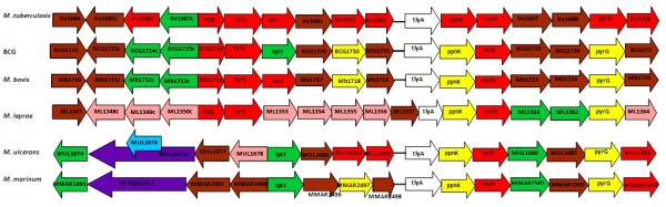
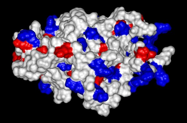
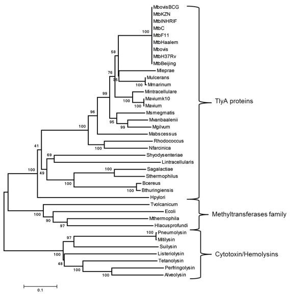
Results: Bioinformatics analysis of TlyA identified a ribosomal protein binding domain (S4 domain), located between residues 5 and 68 as well as an FtsJ-like methyltranferase domain encompassing residues 62 and 247, all of which have been previously described in translation machinery-associated proteins. Subcellular localization prediction showed that TlyA lacks a signal peptide and its hydrophobicity profile showed no evidence of transmembrane helices. These findings suggested that it may not be attached to the membrane, which is consistent with a cytoplasmic localization. Three-dimensional modeling of TlyA showed a consensus structure, having a common core formed by a six-stranded β-sheet between two α-helix layers, which is consistent with an RNA methyltransferase structure. Phylogenetic analyses showed high conservation of the tlyA gene among Mycobacterium species. Additionally, the nucleotide substitution rates suggested purifying selection during tlyA gene evolution and the absence of a common ancestor between TlyA proteins and bacterial pore-forming proteins.

Conclusion: Altogether, our manual in silico curation suggested that TlyA is involved in ribosomal biogenesis and that there is a functional annotation error regarding this protein family in several microbial and plant genomes, including the M. tuberculosis genome.


Figures

Figure 1
Figure 1
Hemolysin A family (InterPro: IPR004538) taxonomic coverage of 864 proteins. (A). Taxonomy-tree nodes are placed on the inner circles and radial lines lead to the description for each node. Proteins annotated by taxonomic division are numbered [http://www.ebi.ac.uk/interpro/IEntry?ac=IPR004538]. (B). Multiple sequence alignment of TlyA protein homologs in Mycobacterium. Sequences are colored according to amino acid physicochemical properties; the catalytic tetrad is marked by black asterisks (K69-D154-K182-E238). Similarity values for each amino acid are represented by bars (pink). Organism names correspond to nomenclature found in the tuberculosis database [http://www.tbdb.org/] (CLC sequence viewer alignment representation).
Figure 2
Figure 2
tlyA transcriptional unit gene syntheny. Genes are represented as arrows and are drawn according to transcriptional orientation and genome functional annotation. Arrow color representation for gene functional annotation is as follows: green for cell wall synthesis, red for signal transduction, brown for evolutionarily conserved, yellow for intermediary metabolism, white for tlyA, pink for pseudogenes, purple for PE/PPE and blue for IS/phages.
Figure 3
Figure 3
Modeled spatial configuration built using UCSF Chimera. Ribbon (A) and space-filling (B) representations of TlyA catalytic tetrad (red), AdoMet binding motif GASTG90-94 (green) and S4 RNA binding domain (blue). Structural model was stored on Protein Model Database [PMDB: PM0076044, http://mi.caspur.it/PMDB/].
Figure 4
Figure 4
Diagram of charge distribution on TlyA Connolly surface. Three-dimensional modeled structure according to positive (blue) and negative (red) charge distribution on the protein surface.
Figure 5
Figure 5
Evolutionary relationships of 34 taxon at amino acid level. Evolutionary relationships were inferred by the NJ method. The optimal tree had 5,628 branch length sum 5,000 replicates were carried out; bootstrap values are shown at each cluster.

Similar articles

Cited by

References

    1. Dorman SE, Chaisson RE. From magic bullets back to the magic mountain: the rise of extensively drug-resistant tuberculosis. Nat Med. 2007;13:295–298. doi: 10.1038/nm0307-295. - DOI - PubMed
    1. Tomioka H. Prospects for development of new antimycobacterial drugs. J Infect Chemother. 2000;6:8–20. doi: 10.1007/s101560050043. - DOI - PubMed
    1. Warner DF, Mizrahi V. Tuberculosis chemotherapy: the influence of bacillary stress and damage response pathways on drug efficacy. Clin Microbiol Rev. 2006;19:558–570. doi: 10.1128/CMR.00060-05. - DOI - PMC - PubMed
    1. Felnagle EA, Rondon MR, Berti AD, Crosby HA, Thomas MG. Identification of the biosynthetic gene cluster and an additional gene for resistance to the antituberculosis drug capreomycin. Appl Environ Microbiol. 2007;73:4162–4170. doi: 10.1128/AEM.00485-07. - DOI - PMC - PubMed
    1. Johansen SK, Maus CE, Plikaytis BB, Douthwaite S. Capreomycin binds across the ribosomal subunit interface using tlyA-encoded 2'-O-methylations in 16S and 23S rRNAs. Mol Cell. 2006;23:173–182. doi: 10.1016/j.molcel.2006.05.044. - DOI - PubMed

MeSH terms

LinkOut - more resources