

In vivo visualization of olfactory pathophysiology induced by intranasal cadmium instillation in mice
- PMID: 21443902
- PMCID: PMC3951954
- DOI: 10.1016/j.neuro.2011.03.007
In vivo visualization of olfactory pathophysiology induced by intranasal cadmium instillation in mice
Abstract
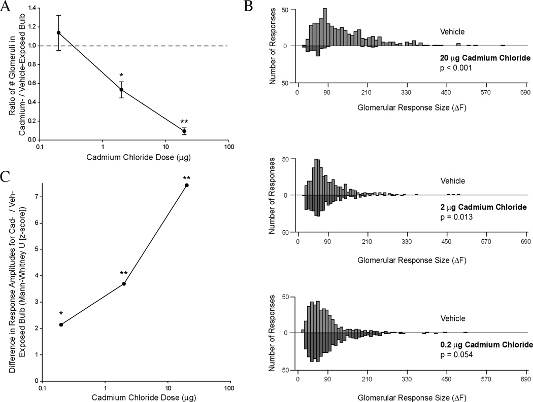
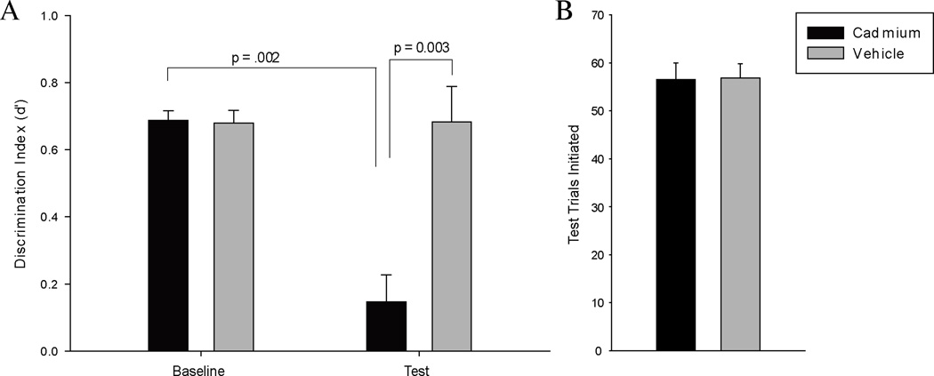
Intranasal exposure to cadmium has been related to olfactory dysfunction in humans and to nasal epithelial damage and altered odorant-guided behavior in rodent models. The pathophysiology underlying these deficits has not been fully elucidated. Here we use optical imaging techniques to visualize odorant-evoked neurotransmitter release from the olfactory nerve into the brain's olfactory bulbs in vivo in mice. Intranasal cadmium chloride instillations reduced this sensory activity by up to 91% in a dose-dependent manner. In the olfactory bulbs, afferents from the olfactory epithelium could be quantified by their expression of a genetically encoded fluorescent marker for olfactory marker protein. At the highest dose tested, cadmium exposure reduced the density of these projections by 20%. In a behavioral psychophysical task, mice were trained to sample from an odor port and make a response when they detected an odorant against a background of room air. After intranasal cadmium exposure, mice were unable to detect the target odor. These experiments serve as proof of concept for a new approach to the study of the neural effects of inhaled toxicants. The use of in vivo functional imaging of the neuronal populations exposed to the toxicant permits the direct observation of primary pathophysiology. In this study optical imaging revealed significant reductions in odorant-evoked release from the olfactory nerve at a cadmium chloride dose two orders of magnitude less than that required to induce morphological changes in the nerve in the same animals, demonstrating that it is a more sensitive technique for assessing the consequences of intranasal neurotoxicant exposure. This approach is potentially useful in exploring the effects of any putative neurotoxicant that can be delivered intranasally.
Copyright © 2011. Published by Elsevier B.V.
Figures


References
-
- Agency for Toxic Substances & Disease Registry. Toxicological profile for cadmium.. Chemical Abstract Service Registry No.7440-43-49. Atlanta, GA: Division of Toxicology and Environmental Medicine; 2008.
-
- Baker H, Genter MB. The Olfactory System and the Nasal Mucosa as Portals of Entry of Viruses, Drugs, and Other Exogenous Agents into the Brain. In: Doty RL, editor. Handbook of 13 Olfaction and Gustation. 2nd edition. New York: Marcel Dekker; 2003. pp. 909–950.
-
- Bondier JR, Michel G, Propper A. Harmful effects of cadmium on olfactory system in mice. Inhalation Toxicology. 2008;20(13):1169–1177. - PubMed
-
- Bozza T, McGann JP, Mombaerts P, Wachowiak M. In vivo imaging of neuronal activity by targeted expression of a genetically encoded probe in the mouse. Neuron. 2004;42:9–21. - PubMed
Publication types
MeSH terms
Substances
Grants and funding
LinkOut - more resources
Full Text Sources
