. 2011 May 15;71(10):3505-15.
doi: 10.1158/0008-5472.CAN-10-4316. Epub 2011 Mar 28.

Tumor-evoked regulatory B cells promote breast cancer metastasis by converting resting CD4⁺ T cells to T-regulatory cells

Affiliations

Tumor-evoked regulatory B cells promote breast cancer metastasis by converting resting CD4⁺ T cells to T-regulatory cells

Purevdorj B Olkhanud et al. Cancer Res. .

Abstract

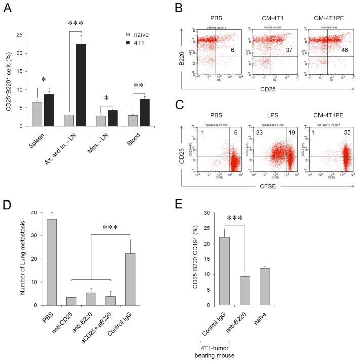
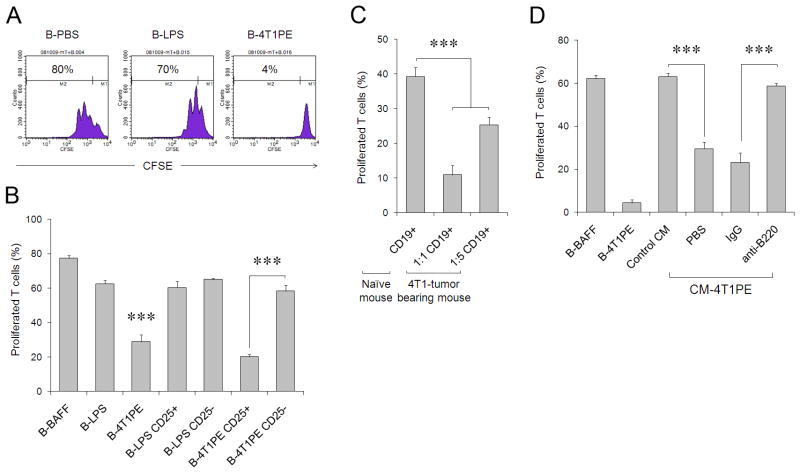
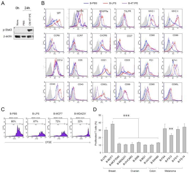
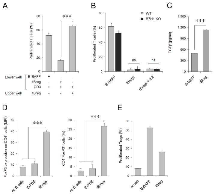
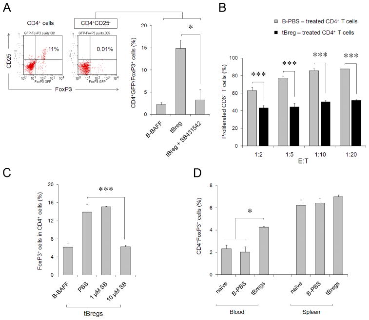
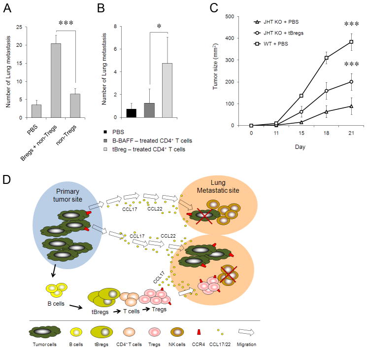
Pulmonary metastasis of breast cancer requires recruitment and expansion of T-regulatory cells (Treg) that promote escape from host protective immune cells. However, it remains unclear precisely how tumors recruit Tregs to support metastatic growth. Here we report the mechanistic involvement of a unique and previously undescribed subset of regulatory B cells. These cells, designated tumor-evoked Bregs (tBreg), phenotypically resemble activated but poorly proliferative mature B2 cells (CD19(+) CD25(High) CD69(High)) that express constitutively active Stat3 and B7-H1(High) CD81(High) CD86(High) CD62L(Low) IgM(Int). Our studies with the mouse 4T1 model of breast cancer indicate that the primary role of tBregs in lung metastases is to induce TGF-β-dependent conversion of FoxP3(+) Tregs from resting CD4(+) T cells. In the absence of tBregs, 4T1 tumors cannot metastasize into the lungs efficiently due to poor Treg conversion. Our findings have important clinical implications, as they suggest that tBregs must be controlled to interrupt the initiation of a key cancer-induced immunosuppressive event that is critical to support cancer metastasis.


Conflict of interest statement

Conflict-of-interest disclosure: The authors work for the US government and declare no competing financial interests.

Figures

FIGURE 1
FIGURE 1
(A) 4T1 cancer -bearing mice (black bars) have a higher proportion of CD25+B220+ cells (% ± SEM of three mice per group) in peripheral blood and secondary lymphoid organs (Ax, axillary; In, inguinal; and Mes, mesenteric) compared with naïve BALB/C mice (grey bars). (B) CM from non-metastatic 4T1-PE cells (CM-4T1PE) had a greater ability to generate CD25+CD19+B220+ cells in vivo, than CM from metastatic 4T1 cells (CM-4T1). Shown is proportion of cells within CD19+ cells. Naïve BALB/C mice were i.p. injected with 0.5 ml CMs or control medium (Mock) once a day four times and splenocytes were stained for CD25 and B220 cells five days after last treatment. (C) Poorly proliferative CD25+B220+ B cells are generated in vitro from naïve mouse B cells after treatment with CM-4T1PE for two days. Control B cells were treated with LPS (B-LPS), or PBS (B-PBS). Histograms show percentage of proliferated (CFSE-diluted) B cells. Numbers are for % of cells in corresponding quadrants. The results in A–C were repeated at least three times. (D, E) B220+CD25+ tBregs are required for lung metastasis. (D) Mean lung metastatic foci ± SEM of four mice per group experiments reproduced three times. 4T1.2 tumor-bearing BALB/C mice were depleted of B220+ and CD25+ cells by i.p. injecting anti-B220 and anti-CD25 Abs alone or together (aCD25+aB220), respectively. Control mice were treated with isotype-matched antibody (IgG). (E) Y-axis shows % ± SEM of CD25+B220+ cells (within CD19+ cells) in spleens of four tumor-bearing and naive mice per group experiments treated with anti-B220 Ab or IgG. From here on, *P<0.05, **P<0.01; *** P<0.001.
FIGURE 2
FIGURE 2
(A) CM-4T1PE, but not mock (B-PBS) or LPS (B-LPS), treated B cells (B-4T1PE) inhibit proliferation of T cells stimulated with anti-CD3/CD28 Abs. B cells and CFSE-labeled T cells (responder) cells were cultured at a 1:1 ratio for four days in the presence of 50 U/ml IL-2. (B) The suppressive activity of B-4T1PE is retained in CD25+ subset. CD25+ and CD25 subsets of B-LPS and B-4T1PE cells were purified using anti-CD25 Ab and tested as in (A). (C) B cells isolated from 4T1 tumor-bearing mice also suppress T cell activity. Splenic CD19+ were isolated from 4T1 tumor-bearing mice and tested as in (A) after mixing with CFSE-labeled T cells in 1:1 and 1:5 B and T cell ratio, while control naïve mouse B cells were used at 1:1 ratio. (D) Splenic CD19+ B cells were isolated from naïve BALB/C mice i.p. injected with control CM or cancer CM (CM-4T1PE, see Fig. 1B) and tested in vitro for the ability to inhibit T cell proliferation as in (A). Purified B cells from CM-4T1PE-treated mice were also depleted using anti-B220 Ab or control IgG prior to mixing with T cells. Controls were B cells in vitro cultured with BAFF (to maintain viability of control cells) or CM-4T1PE (B-BAFF and B-4T1PE, respectively). Shown is % ± SEM of proliferated T cells of triplicates repeated at least three times.
FIGURE 3
FIGURE 3
(A) The CM-4T1PE –treated B cells (tBregs), freshly isolated (None) and PBS-treated B cells (PBS) were lysed and tested for phospho-Stat3 and β-actin using corresponding Abs by western blotting. (B) Surface marker expression (FACS analysis) of purified murine B cells treated with tumor CM (tBregs, pink line), or LPS (B-LPS, red line), or PBS (B-PBS, blue line) after staining with Abs to corresponding surface markers (indicated), or isotype-matched control Ab (grey filled area). (C) Human B cells treated with CM of MDA-MB-231 cells (B-MDA231), but not MCF-7 cells (B-MCF7), or LPS (B-LPS), or PBS (B-PBS), suppress proliferation of T cells. Histograms (B,C) and graphs (D) show % (± SEM of triplicates) of CD3+ T cells that diluted CFSC (proliferated) when mixed with B cells at a 1:1 ratio as in Fig. 2A. (D) Similarly with B-MDA231, B cells treated with CM of other human cancers (depicted on X-axis) also inhibit T cell proliferation. All data were repeated at least three times.
FIGURE 4
FIGURE 4
(A) The suppressive activity of tBregs requires cell contact, as they cannot suppress proliferation of T cells if the cells are physically separated (placed in the upper and lower chambers of trans-well plate, respectively). Controls were B-BAFF and T cells activated with anti-CD3/CD28 Abs (αCD3/CD28). (B) Despite high levels of B7-H1 (Fig. 3B), both w.t. tBregs (grey bars) and B7-H1 deficient tBregs (black bars) can suppress T cells regardless of the presence of exogenously added IL-2 (tBregs+IL2). (C) tBregs secrete high levels of TGFβ1 (pg/ml), as tested by ELISA assay after two days of incubation as in Fig. 1C. (D) tBregs promote Treg conversion in vitro when co-cultured with purified non-Treg CD4+ T cells for five days in the presence anti-CD3/CD28 Abs and 500 U/ml IL-2. Y-axis shows mean fluorescence intensity (MFI, left panel) and percentage (right panel) of FoxP3+ within CD4+ T cells. (E) Purified and CFSE-labeled Tregs (CD25+CD4+) were stimulated with anti-CD3/CD28 Abs and IL-2 (500 U/ml) in the presence of tBregs or control B cells. Y-axis shows % of proliferated (CFSE diluted) cells ± SEM of triplicate experiments. The experiments were reproduced at least three times.
FIGURE 5
FIGURE 5
(A) Purified GFPCD4+ T cells from FoxP3-GFP were depleted of CD25+ cells (< 0.01%, right dot blot, inlet) and stimulated with anti-CD3/CD28 Abs and IL-2 (500 U/ml) in the presence of tBregs or control B cells. Y-axis shows % ± SEM of GFP+(FoxP3+)Tregs after 5 days of culture of a triplicate assay. Left dot blot (inlet) shows proportion of GFP+CD25+ of purified CD4+ T cells before CD25 depletion. (B) The converted FoxP3+ T cells (as in Fig. 4D) are Tregs, as they (after two rounds of depletion of B cells, >98% pure CD4+ cells) suppressed proliferation of CFSE-labeled CD8+ T cells stimulated with anti-CD3/CD28 Abs and 500 U/ml IL-2 at the indicated E:T ratio. Y-axis shows percentage of proliferated CFSE-labeled CD8+ cells of a triplicate experiment repeated twice. P values are for comparisons between tBreg- (black bars) and normal B cell- (grey bars) treated CD4+ T cells. tBreg-mediated Treg conversion from non-Tregs (% of GFP+CD4+ cells in A or FoxP3+CD4+ cells in C, Y-axis) requires TGFβ signaling, as it was blocked with 10 μM SB431542. (D) Unlike B-PBS, tBregs expand FoxP3+CD4+ T cells in vivo. Three naïve BALB/C mice per group were i.p. injected with 107 B cells and the proportion of FoxP3+CD4+ T cells (% ± SEM, Y-axis) was evaluated after 5 days in the blood and spleens by FACS. All data shown were reproduced at least three times.
FIGURE 6
FIGURE 6
(AB) tBregs support lung metastases via Treg generation. The inability of 4T1.2 cells to metastasize in T and B cell deficient NOD/SCID mice is reversed by transfer of tBregs together with non-Tregs (A) or newly tBreg-generated Tregs (B, depleted of B cells as in Fig. 4D). Control mice received CD25CD4+ T cells (non-Tregs, A) alone, or tBregs alone (not shown), or T cells cultured with mock-treated B cells (B). (C) Poor growth of s.c. challenged B16 melanoma in mice deficient in mature B cells (JHT KO) can be reversed by adoptive transfer of congeneic tBregs (splenic B cells from naïve C57BL/6 mice treated with CM-4T1PE as in Fig. 1C). Shown, mean lung metastatic foci (A,B) or tumor size (C) ± SEM of four-five mice per group experiments reproduced three times. (D) Summary schema which expands our recent report on the importance of Tregs in lung metastases (3) by adding a “missing link”, tBregs. Our data indicate that as long as cancer persists, it induces the generation of tBregs from resting B cells by producing yet to be identified soluble factors. As a result, tBregs induce TGFβ-dependent FoxP3+ Treg conversion of non-Treg T cells and thereby promote lung metastasis by presumably utilizing the same mechanism we previously reported, such as by migrating into CCL17/CCL22-producing lungs to directly kill antitumor NK cells (3). In the absence of tBregs, cancer cannot metastasize into the lungs due to a poor Treg conversion.

References

    1. Youn JI, Nagaraj S, Collazo M, Gabrilovich DI. Subsets of myeloid-derived suppressor cells in tumor-bearing mice. J Immunol. 2008;181:5791–802. - PMC - PubMed
    1. Liu VC, Wong LY, Jang T, Shah AH, Park I, Yang X, et al. Tumor evasion of the immune system by converting CD4+ J Immunol. 2007;178:2883–92. - PubMed
    1. Olkhanud PB, Baatar D, Bodogai M, Hakim F, Gress R, Anderson RL, et al. Breast cancer lung metastasis requires expression of chemokine receptor CCR4 and regulatory T cells. Cancer Res. 2009;69:5996–6004. - PMC - PubMed
    1. Lelekakis M, Moseley JM, Martin TJ, Hards D, Williams E, Ho P, et al. A novel orthotopic model of breast cancer metastasis to bone. Clin Exp Metastasis. 1999;17:163–70. - PubMed
    1. DuPre SA, Redelman D, Hunter KW., Jr The mouse mammary carcinoma 4T1: characterization of the cellular landscape of primary tumours and metastatic tumour foci. Int J Exp Pathol. 2007;88:351–60. - PMC - PubMed

Publication types

Substances