. 2011 Apr 4;193(1):31-9.
doi: 10.1083/jcb.201011110. Epub 2011 Mar 28.

Paraspeckles are subpopulation-specific nuclear bodies that are not essential in mice

Affiliations

Paraspeckles are subpopulation-specific nuclear bodies that are not essential in mice

Shinichi Nakagawa et al. J Cell Biol. .

Abstract

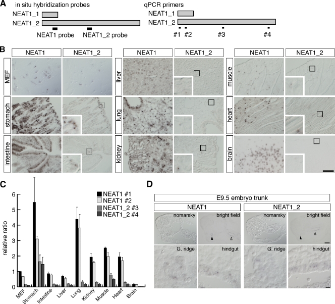
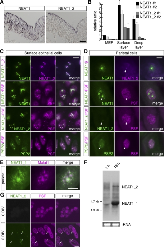
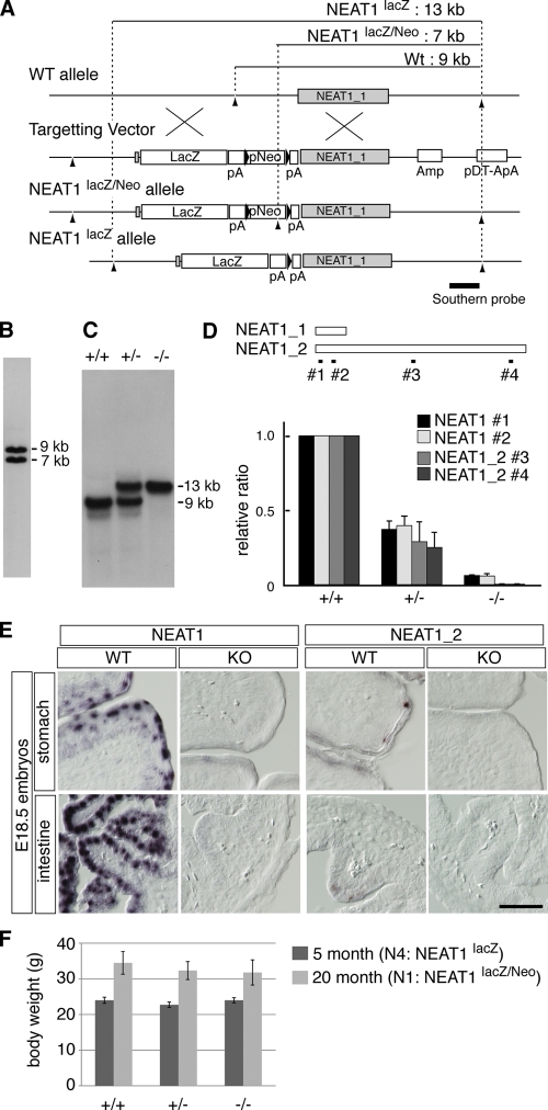
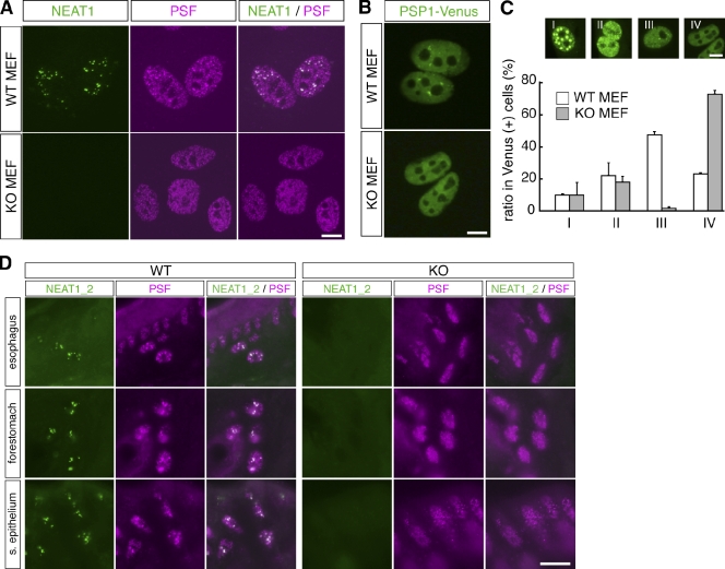
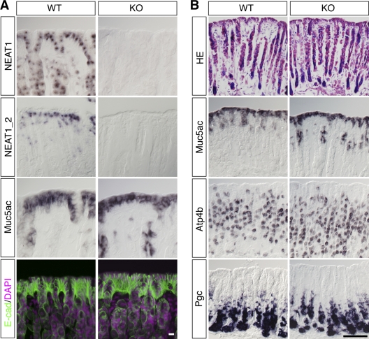
Nuclei of higher organisms are well structured and have multiple, distinct nuclear compartments or nuclear bodies. Paraspeckles are recently identified mammal-specific nuclear bodies ubiquitously found in most cells cultured in vitro. To investigate the physiological role of paraspeckles, we examined the in vivo expression patterns of two long noncoding RNAs, NEAT1_1 and NEAT1_2, which are essential for the architectural integrity of nuclear bodies. Unexpectedly, these genes were only strongly expressed in a particular subpopulation of cells in adult mouse tissues, and prominent paraspeckle formation was observed only in the cells highly expressing NEAT1_2. To further investigate the cellular functions of paraspeckles, we created an animal model lacking NEAT1 by gene targeting. These knockout mice were viable and fertile under laboratory growth conditions, showing no apparent phenotypes except for the disappearance of paraspeckles. We propose that paraspeckles are nonessential, subpopulation-specific nuclear bodies formed secondary to particular environmental triggers.


Figures

Figure 1.
Figure 1.
NEAT1_1 and NEAT1_2 are expressed in a particular subpopulation of cells in mouse tissues. (A) Schematic drawing of the position of the probes used for in situ hybridization and primers used for qPCR analysis. (B) Expression of NEAT1 and NEAT1_2 in various adult organs detected by in situ hybridization. Insets show higher magnification images of the region indicated by boxes. (C) qPCR analysis of expression of NEAT1 and NEAT1_2. (D) Expression of NEAT1 and NEAT1_2 in the trunk region of E9.5 embryos. Faint expression is observed in cells in the hindgut (open arrowheads) and genital ridges (arrowheads). Bars, 100 µm.
Figure 2.
Figure 2.
Paraspeckles are formed only in a small subpopulation of cells expressing NEAT1_2. (A) Expression pattern of NEAT1 and NEAT1_2 in the zymogenic region of the adult stomach detected by in situ hybridization. NEAT1_2 expression is restricted to the surface epithelial cells facing the lumen of the stomach. (B) qPCR analysis of the expression of NEAT1 and NEAT1_2 in the dissected surface and deep layer of the gastric epithelium. (C and D) Subnuclear distribution of NEAT1, NEAT1_2 detected by FISH, and the paraspeckle marker PSF and PSP2 in the surface epithelial cells (C) and parietal cells (D). Arrowheads in D indicate the putative transcription sites of NEAT1_2. (E) Different expression of NEAT1_1 and Malat1 in parietal cells that lack expression of NEAT1_2. (F) Induction of NEAT1 expression in cultured MEFs. (G) Paraspeckle formation is rapidly induced in MEFs cultured in vitro (DIV, days in vitro). Bars: (A) 100 µm; (C, D, and G) 10 µm; (E) 5 µm.
Figure 3.
Figure 3.
Generation of NEAT1 knockout mouse. (A) Targeting strategy for disruption of the NEAT1 gene. Arrowheads indicate SphI restriction sites, and black triangles indicate the FRT sequences flanking the Neo cassette. (B) Southern blot analysis of SphI-digested DNA from embryonic stem cells that underwent homologous recombination. (C) Southern blot analysis of SphI-digested tail DNA from of NEAT1lacZ mice. (D) Quantitative PCR analysis of NEAT1 and NEAT1_2 transcripts. (E) In situ hybridization of NEAT1 and NEAT1_2 in stomach and intestine of E18.5 embryos. Bar, 100 µm. (F) Body weight of female littermates (n = 3 for each genotype).
Figure 4.
Figure 4.
Paraspeckles are not formed in NEAT1 knockout mice. (A) Subnuclear localization of NEAT1_2 and the paraspeckle marker PSF in MEFs. The foci of PSF were not observable in MEFs from NEAT1 knockout mice. (B) Typical distribution of PSP1-Venus in MEFs from wild-type (WT) and knockout (KO) mice. The paraspeckle marker accumulated as discrete foci in WT MEFs, but not in knockout MEFs. (C) Quantitative analysis of subnuclear localization of PSP1-Venus in WT and knockout MEFs. Overexpressed PSP1-Venus signals were categorized as types I–IV. Note that typical paraspeckle-like distribution (type III) was rarely observed in knockout MEFs. (D) Loss of punctate signals of paraspeckle marker PSF in epithelial cells of esophagus, forestomach, and surface epithelium (s. epithelium) of zymogenic stomach in the knockout mice. Bars, 10 µm.
Figure 5.
Figure 5.
Normal tissue organization of stomach in NEAT1 knockout mice. (A) Expression pattern of NEAT1, NEAT1_2, surface epithelial cell marker Muc5ac, and cell–cell junction marker E-cadherin (E-cad) in the zymogenic epithelium in the wild-type and NEAT1 knockout stomach. (B) Normal organization of tissue integrity revealed by histological HE staining and in situ hybridization for Muc5ac, Atp4b, and Pgc, which are expressed in surface mucosa cells, acid-producing parietal cells, and enzyme-producing zymogenic cells, respectively. Bars: (A) 10 µm; (B) 100 µm.

References

    1. Bond C.S., Fox A.H. 2009. Paraspeckles: nuclear bodies built on long noncoding RNA. J. Cell Biol. 186:637–644 10.1083/jcb.200906113 - DOI - PMC - PubMed
    1. Chen L.-L., Carmichael G.G. 2009. Altered nuclear retention of mRNAs containing inverted repeats in human embryonic stem cells: functional role of a nuclear noncoding RNA. Mol. Cell. 35:467–478 10.1016/j.molcel.2009.06.027 - DOI - PMC - PubMed
    1. Chen L.L., Carmichael G.G. 2010. Decoding the function of nuclear long non-coding RNAs. Curr. Opin. Cell Biol. 22:357–364 10.1016/j.ceb.2010.03.003 - DOI - PMC - PubMed
    1. Chen L.L., DeCerbo J.N., Carmichael G.G. 2008. Alu element-mediated gene silencing. EMBO J. 27:1694–1705 10.1038/emboj.2008.94 - DOI - PMC - PubMed
    1. Clemson C.M., Hutchinson J.N., Sara S.A., Ensminger A.W., Fox A.H., Chess A., Lawrence J.B. 2009. An architectural role for a nuclear noncoding RNA: NEAT1 RNA is essential for the structure of paraspeckles. Mol. Cell. 33:717–726 10.1016/j.molcel.2009.01.026 - DOI - PMC - PubMed

Publication types

Substances