. 2011 Apr 4;193(1):97-108.
doi: 10.1083/jcb.201011083. Epub 2011 Mar 28.

Replication stress induces 53BP1-containing OPT domains in G1 cells

Affiliations

Replication stress induces 53BP1-containing OPT domains in G1 cells

Jeanine A Harrigan et al. J Cell Biol. .

Abstract

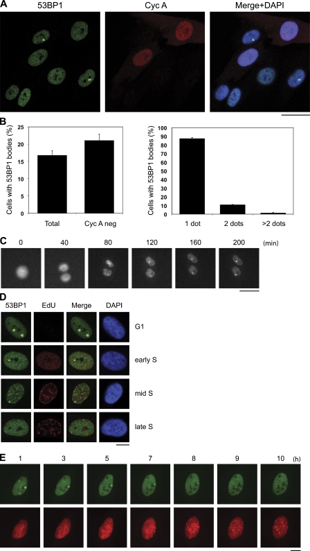
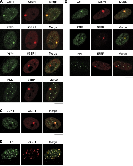
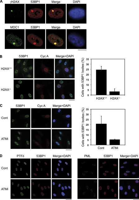
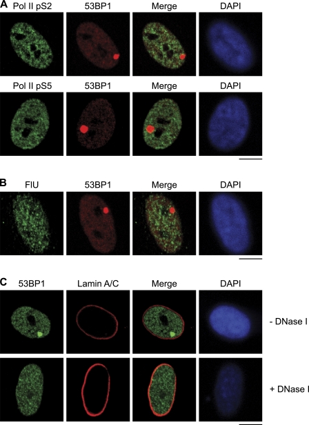
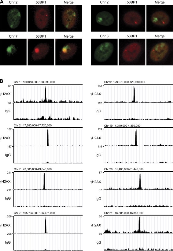
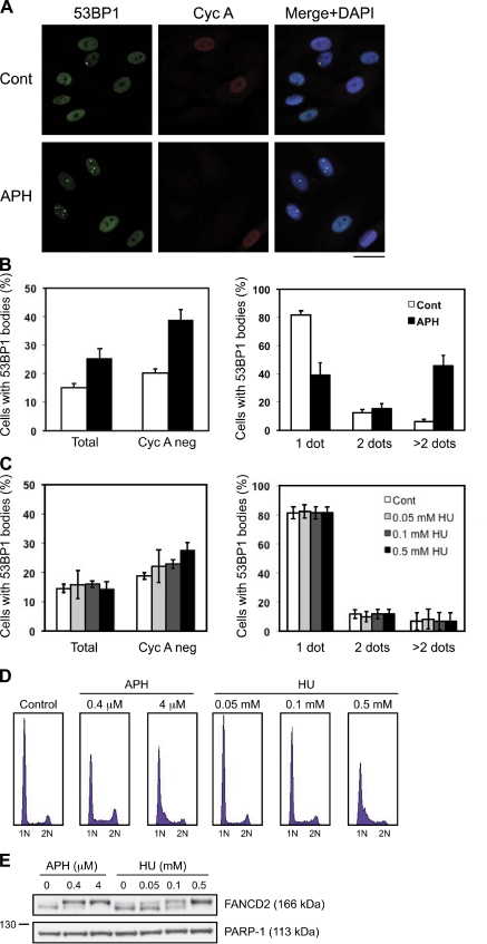
Chromosomal deletions and rearrangements in tumors are often associated with common fragile sites, which are specific genomic loci prone to gaps and breaks in metaphase chromosomes. Common fragile sites appear to arise through incomplete DNA replication because they are induced after partial replication inhibition by agents such as aphidicolin. Here, we show that in G1 cells, large nuclear bodies arise that contain p53 binding protein 1 (53BP1), phosphorylated H2AX (γH2AX), and mediator of DNA damage checkpoint 1 (MDC1), as well as components of previously characterized OPT (Oct-1, PTF, transcription) domains. Notably, we find that incubating cells with low aphidicolin doses increases the incidence and number of 53BP1-OPT domains in G1 cells, and by chromatin immunoprecipitation and massively parallel sequencing analysis of γH2AX, we demonstrate that OPT domains are enriched at common fragile sites. These findings invoke a model wherein incomplete DNA synthesis during S phase leads to a DNA damage response and formation of 53BP1-OPT domains in the subsequent G1.


Figures

Figure 1.
Figure 1.
53BP1 accumulates in nuclear bodies in a subset of G1 cells. (A) Asynchronously growing BJ hTERT cells were fixed, and immunofluorescence was performed with mouse anti-53BP1 and rabbit anti–Cyc A antibodies. (B, left) Quantitation of total or Cyc A–negative cells from A that contained 53BP1 nuclear bodies. Results represent the mean ± SD from three experiments (total, n = 4,944; Cyc A negative, n = 3,956). (B, right) Quantitation of Cyc A–negative cells from A that contained 53BP1 bodies. Results represent the mean ± SD from three experiments (n = 829). We note that 53BP1 foci were also observed in 10.8% (±2.8%) of Cyc A–positive cells (not depicted), which likely represent early-to-mid S phase cells (see D). (C) Live cell imaging of U2OS cells stably expressing EGFP-53BP1 from mitosis into G1. (D) BJ hTERT cells were pulsed with EdU for 10 min, fixed, and stained with rabbit anti-53BP1 antibodies. EdU was visualized using click chemistry. (E) Live cell imaging of U2OS cells stably expressing EGFP-53BP1 and mRuby-PCNA from G1 into S phase. Bars: (A and C) 30 µm; (D and E) 10 µm.
Figure 2.
Figure 2.
53BP1 colocalizes with components of OPT domains. (A) Immunofluorescence was performed in BJ primary or BJ hTERT (PTFγ) fibroblasts with mouse anti-53BP1 and rabbit anti–Oct-1, anti-PTFδ, or anti-PTFγ, and rabbit anti-53BP1 and mouse anti-PML antibodies as indicated. Analysis of >100 cells revealed that 88% of 53BP1 nuclear bodies contained at least one PML body. (B) Experiments were performed as in A with U2OS cells and the indicated antibodies. (C) Immunofluorescence was performed in BJ primary fibroblasts with mouse anti-53BP1 and rabbit anti-DDX1 antibodies. (D) BJ hTERT fibroblasts were exposed to 1.5 Gy of ionizing radiation and fixed 1 h later. Immunofluorescence was performed with rabbit anti-PTFδ and mouse anti-53BP1 antibodies. Bars, 10 µm.
Figure 3.
Figure 3.
53BP1-OPT domains represent sites of DNA damage. (A) Immunofluorescence was performed in BJ hTERT fibroblasts with rabbit anti-53BP1 and mouse anti-γH2AX (top) or mouse anti-53BP1 and rabbit anti-MDC1 pSDTD (bottom) antibodies. (B, left) Immunofluorescence was performed in H2AX+/+ or H2AX−/− MEFs with rabbit anti-53BP1 or mouse anti–Cyc A antibodies. (B, right) Quantitation of Cyc A–negative cells that contained 53BP1 nuclear bodies. Results represent the mean ± SD from three experiments (H2AX+/+, n = 636; H2AX−/−, n = 384). (C, left) BJ hTERT fibroblasts were incubated in the absence (Cont) or presence of ATM inhibitor KU55933 (ATMi; 20 µM) for 3 h. Immunofluorescence was performed with mouse anti-53BP1 and rabbit anti–Cyc A antibodies. (C, right) Quantitation of Cyc A–negative cells that contained 53BP1 nuclear bodies. Results represent the mean ± SD from three experiments (Cont, n = 522; ATMi, n = 491). (D) Experiments were performed as in C and immunofluorescence was performed with rabbit anti-PTFδ and mouse anti-53BP1 antibodies (left) or mouse anti-PML and rabbit anti-53BP1 antibodies (right). Bars: (A) 10 µm; (B–D) 30 µm.
Figure 4.
Figure 4.
53BP1-OPT domains do not associate with regions of active transcription and are dependent on DNA. (A) Immunofluorescence was performed in BJ primary fibroblasts with mouse anti-53BP1 and rabbit anti-Pol II pS2 (top) or rabbit anti-53BP1 and mouse anti-Pol II pS5 (bottom) antibodies. (B) BJ primary fibroblasts were incubated for 60 min with FlU and immunofluorescence was performed with rabbit anti-53BP1 and mouse anti-BrdU antibodies. (C) BJ primary fibroblasts were incubated in the absence or presence of DNase I as indicated for 10 min. Immunofluorescence was performed with rabbit anti-53BP1 or mouse anti–Lamin A/C antibodies. DAPI and Lamin A/C were positive and negative controls, respectively. Bars, 10 µm.
Figure 5.
Figure 5.
γH2AX accumulates at discrete regions of the genome in untreated cells. (A) DNA Immuno-FISH was performed in BJ hTERT fibroblasts with anti-53BP1 antibodies and whole chromosome paints. (A, left) Colocalization between 53BP1 nuclear bodies and chromosome regions. (A, right) No colocalization between 53BP1 nuclear bodies and chromosome regions. Bar, 10 µm. (B) ChIP-seq experiments were performed with serum-starved (G0) BJ hTERT fibroblasts and γH2AX antibodies. The eight highest peaks for γH2AX enrichment are displayed along with the negative control (IgG). Data are shown as custom tracks (bin 20) on the UCSC genome browser (data correspond to the Feb. 2009 GRCh37/hg19 genome assembly). The axis scales are presented on the left and chromosome position at the top.
Figure 6.
Figure 6.
Replication perturbation by APH promotes formation of 53BP1-OPT domains. (A) BJ hTERT fibroblasts were incubated in the absence (Cont) or presence of APH (0.4 µM, 24 h) as indicated. Immunofluorescence was performed using mouse anti-53BP1 and rabbit anti–Cyc A antibodies. Bar, 30 µm. (B, left) Quantitation of total or Cyc A–negative cells from A that contained 53BP1 nuclear bodies. Results represent the mean ± SD from three experiments (total: Cont, n = 1,329; APH, n = 1,291; Cyc A negative: Cont, n = 987; APH, n = 843). (B, right) Quantitation of Cyc A–negative cells from A that contained 53BP1 nuclear bodies. Results represent the mean ± SD from three experiments (Cont, n = 199; APH, n = 328). (C) BJ hTERT fibroblasts were incubated in the absence (Cont) or presence of HU for 24 h as indicated. Immunofluorescence was performed as described in A and quantified as in B. Results represent the mean ± SD from three experiments. (D) FACS analysis of cells incubated in the absence or presence of APH or HU as indicated. (E) Cellular lysates from cells treated with APH or HU as in A and C were separated by SDS-PAGE, and Western blotting was performed with antibodies against FANCD2 and PARP-1 (loading control).

References

    1. Ahel D., Horejsí Z., Wiechens N., Polo S.E., Garcia-Wilson E., Ahel I., Flynn H., Skehel M., West S.C., Jackson S.P., et al. 2009. Poly(ADP-ribose)-dependent regulation of DNA repair by the chromatin remodeling enzyme ALC1. Science. 325:1240–1243 10.1126/science.1177321 - DOI - PMC - PubMed
    1. Bouwman P., Aly A., Escandell J.M., Pieterse M., Bartkova J., van der Gulden H., Hiddingh S., Thanasoula M., Kulkarni A., Yang Q., et al. 2010. 53BP1 loss rescues BRCA1 deficiency and is associated with triple-negative and BRCA-mutated breast cancers. Nat. Struct. Mol. Biol. 17:688–695 10.1038/nsmb.1831 - DOI - PMC - PubMed
    1. Branzei D., Foiani M. 2010. Maintaining genome stability at the replication fork. Nat. Rev. Mol. Cell Biol. 11:208–219 10.1038/nrm2852 - DOI - PubMed
    1. Bunting S.F., Callén E., Wong N., Chen H.T., Polato F., Gunn A., Bothmer A., Feldhahn N., Fernandez-Capetillo O., Cao L., et al. 2010. 53BP1 inhibits homologous recombination in Brca1-deficient cells by blocking resection of DNA breaks. Cell. 141:243–254 10.1016/j.cell.2010.03.012 - DOI - PMC - PubMed
    1. Celeste A., Fernandez-Capetillo O., Kruhlak M.J., Pilch D.R., Staudt D.W., Lee A., Bonner R.F., Bonner W.M., Nussenzweig A. 2003. Histone H2AX phosphorylation is dispensable for the initial recognition of DNA breaks. Nat. Cell Biol. 5:675–679 10.1038/ncb1004 - DOI - PubMed

Publication types

MeSH terms

Substances