. 2011 Apr 4;193(1):41-50.
doi: 10.1083/jcb.201008071. Epub 2011 Mar 28.

Attenuation of yeast UPR is essential for survival and is mediated by IRE1 kinase

Affiliations

Attenuation of yeast UPR is essential for survival and is mediated by IRE1 kinase

Aditi Chawla et al. J Cell Biol. .

Abstract

The unfolded protein response (UPR) activates Ire1, an endoplasmic reticulum (ER) resident transmembrane kinase and ribonuclease (RNase), in response to ER stress. We used an in vivo assay, in which disappearance of the UPR-induced spliced HAC1 messenger ribonucleic acid (mRNA) correlates with the recovery of the ER protein-folding capacity, to investigate the attenuation of the UPR in yeast. We find that, once activated, spliced HAC1 mRNA is sustained in cells expressing Ire1 carrying phosphomimetic mutations within the kinase activation loop, suggesting that dephosphorylation of Ire1 is an important step in RNase deactivation. Additionally, spliced HAC1 mRNA is also sustained after UPR induction in cells expressing Ire1 with mutations in the conserved DFG kinase motif (D828A) or a conserved residue (F842) within the activation loop. The importance of proper Ire1 RNase attenuation is demonstrated by the inability of cells expressing Ire1-D828A to grow under ER stress. We propose that the activity of the Ire1 kinase domain plays a role in attenuating its RNase activity when ER function is recovered.


Figures
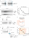
Figure 1.
Figure 1.
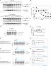
Attenuation of spliced HAC1 mRNA levels during the UPR. (A) ire1Δ yeast strain carrying WT IRE1 (Ire1-WT) was induced for ER stress with tunicamycin (Tm) at either middle (I) or early (II) log phases. Activation of IRE1 was monitored by HAC1 mRNA splicing on the Northern blots of isolated RNA from cells collected at various time points. Positions of spliced and unspliced HAC1 mRNA are indicated. Differences in HAC1 mRNA splicing are highlighted by the dotted blue boxes. (B) An extended UPR time course of Ire1-WT cells in the presence of Tm throughout the time course. The HAC1 mRNA membrane was reprobed for KAR2/BiP mRNA. (C) The percentage of HAC1 splicing (dotted line) over total HAC1 mRNA. (D) Ire1-WT cells continuously incubated with Tm were not able to splice HAC1 mRNA upon addition of 1 µg/ml of fresh Tm at the 8-h time point. (E) Tm in media that were incubated for 8 h was able to splice HAC1 in fresh cells that have never been treated with Tm. (F) The vacuolar carboxypeptidase Y (CPY) activity in Ire1-WT or ire1Δ cells with pRS341 was examined by the ability of CPY to cleave its substrate BTPNA to release p-nitroaniline, which was measured at 410 nm. The value of Ire1-WT cells before the UPR was set to 100% CPY activity, and the percentage of CPY activity in Ire1-WT or ire1Δ cells treated with Tm for the indicated lengths of time was calculated. (G) Levels of CPY activity for Ire1-WT cells were also shown (orange) and superimposed on the graph of HAC1 mRNA splicing from C. Error bars represent standard deviations calculated from at least three independent time courses.
Figure 2.
Figure 2.
Removal of Tm from the medium makes the recovery of spliced HAC1 mRNA faster in Ire1-WT cells but not in Ire1-S840D/S841D/T844D cells. (A) Ire1-WT cells incubated with Tm for the amount of time indicated. (B) Quantitation of HAC1 mRNA splicing (closed square). (C) After a 2-h incubation with Tm, Ire1-WT cells were recovered from ER stress by washing them in warm fresh media (Wash) and incubated further without Tm for the remaining time. The white line between lanes 2 and 3 indicates the removal of intervening lanes. (D) Quantitation of HAC1 mRNA splicing for panel C is shown. Blue numbers represent the amount of time after washing. (E–J) Cells were recovered after 1.5 (E and F)-, 1 (G and H)-, and 0.6 (I and J)-h Tm incubation, and the splicing of HAC1 mRNA was quantified. In I, intervening lanes were spliced out between lanes 2 and 3, lanes 3 and 4, and lanes 4 and 5. (K) Ire1-S840D/S841D/T844D cells continue to retain spliced HAC1 mRNA once activated without recovery. (L) The percentage of spliced HAC1 mRNA (closed circle). (M–P) Ire1-S840D/S841D/T844D cells were recovered from Tm after 2 (M and N)- or 6-h (O and P) incubation, and the percentage of HAC1 mRNA splicing was quantified. In M, the splicing of two different parts of a northern membrane is indicated by the white line between lanes 1 and 2. Error bars indicate standard deviations from at least three independent experiments.
Figure 3.
Figure 3.
Spliced HAC1 mRNA is sustained in Ire1-D828A cells upon ER stress. (A) Ire1-D828A cells were treated continuously with Tm for the indicated length of time for HAC1 mRNA splicing on Northern gels and reprobed for the level of KAR2 mRNA. As a comparison, the Tm time course of Ire1-WT cells (Fig 1B) was shown. (B) The percentage of HAC1 mRNA spliced for Ire1-WT (closed square) and Ire1-D828A (closed circle). (C–J) Sustained presence of spliced HAC1 RNA in Ire1-D828A cells even after the removal of Tm and resuspension in fresh media without Tm. (D) The percentage of HAC1 mRNA splicing in cells treated with continuous Tm (closed circle) and in cells recovered after 2-h Tm treatment (open circle). (F, H, and J) Close up of the quantitation for the first 5 h of recovery. Error bars are standard deviations from at least three repeats.
Figure 4.
Figure 4.
An additional kinase domain mutation displays a sustained HAC1 mRNA-splicing phenotype, and the inability to attenuate HAC1 splicing in Ire1-D828A is more sensitive to ER stress. (A) The D828A mutation partially rescues RNase-inactive Ire1-S840A/S841A/T844A. For each mutant, 0- and 2-h time points are shown. (B) Alignment of amino acid residues in the activation loop surrounding the DFG motif (shown in red). Serines at 840 and 841 and threonine at 844 (shown in blue) become phosphorylated upon UPR activation (Shamu and Walter, 1996; Lee et al., 2008). The conserved phenylalanine (F843) is shown in the purple box. (C) A mutation at the conserved phenylalanine in the activation loop (Ire1-F842A) phenocopies the Ire1-D828A mutant. (D) Ire1-D828A cells are more sensitive to ER stress. Ire1-WT or Ire1-D828A cells were grown to an OD600nm of 0.25, and then cells were serially diluted by fivefold and spotted on plates containing different amounts of Tm (0, 0.1, and 0.4 µg/ml). Error bars represent standard deviations from three independent experiments. Dros., Drosophila melanogaster.

Comment in

References

    1. Adams J.A., Taylor S.S. 1992. Energetic limits of phosphotransfer in the catalytic subunit of cAMP-dependent protein kinase as measured by viscosity experiments. Biochemistry. 31:8516–8522 10.1021/bi00151a019 - DOI - PubMed
    1. Aragón T., van Anken E., Pincus D., Serafimova I.M., Korennykh A.V., Rubio C.A., Walter P. 2009. Messenger RNA targeting to endoplasmic reticulum stress signalling sites. Nature. 457:736–740 10.1038/nature07641 - DOI - PMC - PubMed
    1. Cox J.S., Walter P. 1996. A novel mechanism for regulating activity of a transcription factor that controls the unfolded protein response. Cell. 87:391–404 10.1016/S0092-8674(00)81360-4 - DOI - PubMed
    1. Cox J.S., Shamu C.E., Walter P. 1993. Transcriptional induction of genes encoding endoplasmic reticulum resident proteins requires a transmembrane protein kinase. Cell. 73:1197–1206 10.1016/0092-8674(93)90648-A - DOI - PubMed
    1. Credle J.J., Finer-Moore J.S., Papa F.R., Stroud R.M., Walter P. 2005. On the mechanism of sensing unfolded protein in the endoplasmic reticulum. Proc. Natl. Acad. Sci. USA. 102:18773–18784 10.1073/pnas.0509487102 - DOI - PMC - PubMed

Publication types

MeSH terms

Substances