. 2011 May;31(10):2040-52.
doi: 10.1128/MCB.01437-10. Epub 2011 Mar 28.

Distinct mechanisms of ferritin delivery to lysosomes in iron-depleted and iron-replete cells

Affiliations

Distinct mechanisms of ferritin delivery to lysosomes in iron-depleted and iron-replete cells

Takeshi Asano et al. Mol Cell Biol. 2011 May.

Abstract

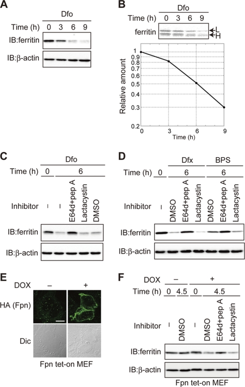
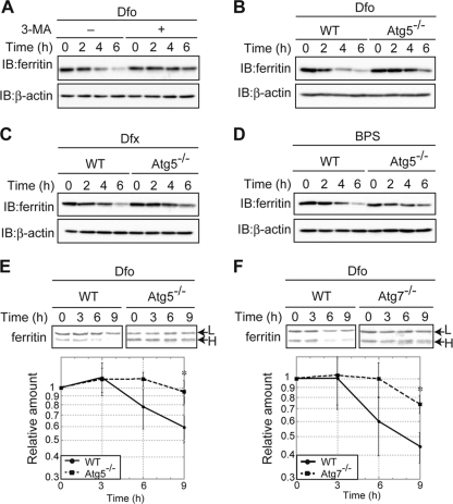
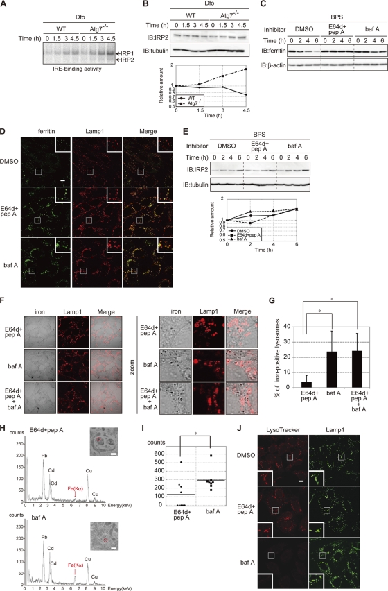
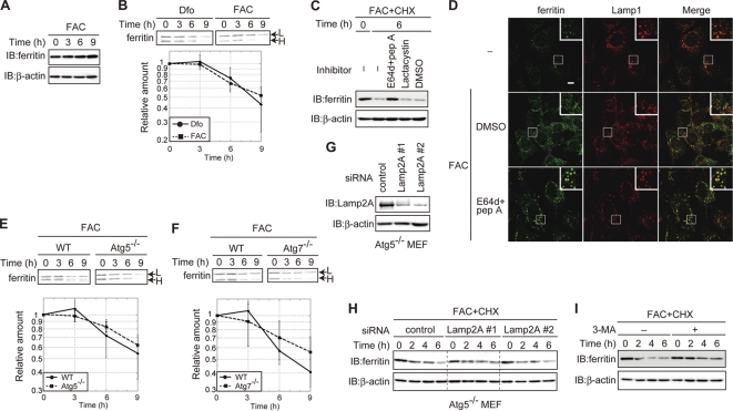
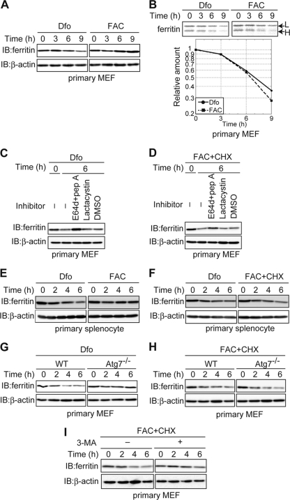
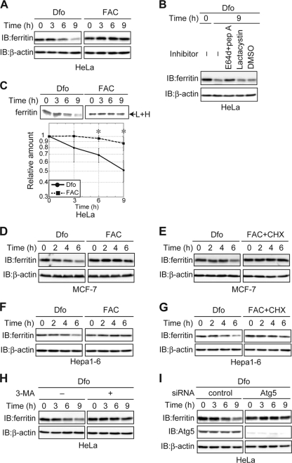
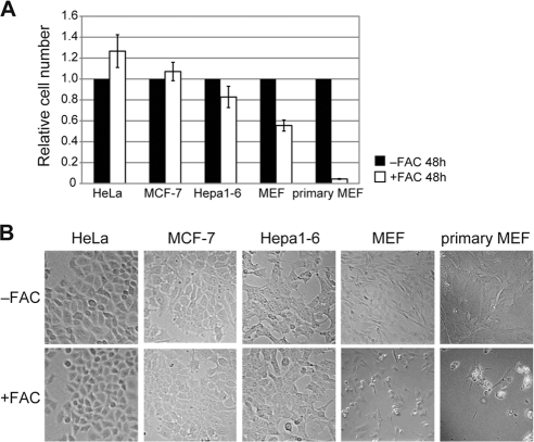
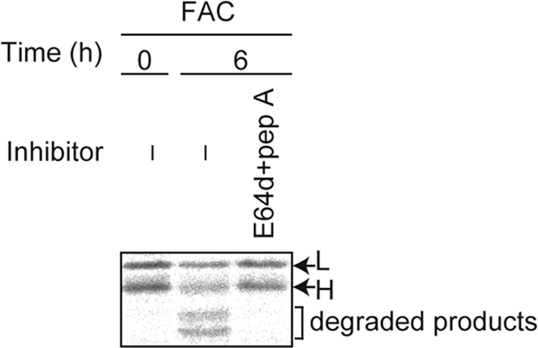
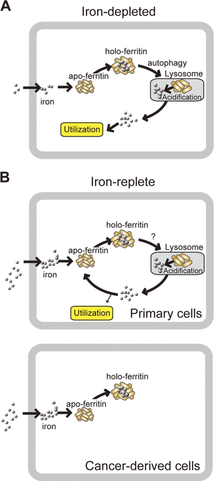
Ferritin is a cytosolic protein that stores excess iron, thereby protecting cells from iron toxicity. Ferritin-stored iron is believed to be utilized when cells become iron deficient; however, the mechanisms underlying the extraction of iron from ferritin have yet to be fully elucidated. Here, we demonstrate that ferritin is degraded in the lysosome under iron-depleted conditions and that the acidic environment of the lysosome is crucial for iron extraction from ferritin and utilization by cells. Ferritin was targeted for degradation in the lysosome even under iron-replete conditions in primary cells; however, the mechanisms underlying lysosomal targeting of ferritin were distinct under depleted and replete conditions. In iron-depleted cells, ferritin was targeted to the lysosome via a mechanism that involved autophagy. In contrast, lysosomal targeting of ferritin in iron-replete cells did not involve autophagy. The autophagy-independent pathway of ferritin delivery to lysosomes was deficient in several cancer-derived cells, and cancer-derived cell lines are more resistant to iron toxicity than primary cells. Collectively, these results suggest that ferritin trafficking may be differentially regulated by cell type and that loss of ferritin delivery to the lysosome under iron-replete conditions may be related to oncogenic cellular transformation.


Figures

Fig. 1.
Fig. 1.
Ferritin is degraded via the lysosome under iron-starved conditions. (A) Decreased levels of ferritin in Dfo-treated MEFs. MEFs pretreated with FAC were cultured with Dfo for the indicated times. Cell lysates were analyzed by immunoblotting (IB) with antiferritin or anti-β-actin. (B) Ferritin is degraded in Dfo-treated cells. MEFs pretreated with FAC were pulse-labeled with EXPRE35S35S in the presence of Dfo. Cells were cultured in medium containing Dfo and harvested at the indicated times. Antiferritin immunoprecipitates were visualized with a BAS 5000 imager. The upper band is the ferritin L chain (L), and the lower band is the ferritin H chain (H). The amount of ferritin L chain was quantified. (C, D) Ferritin is degraded in the lysosome in MEFs treated with iron chelators. MEFs pretreated with FAC were cultured in the presence of indicated inhibitor(s) or dimethyl sulfoxide (DMSO) as the vehicle control, together with Dfo (C) or Dfx or BPS (D) for 6 h. Cell lysates were analyzed as described for panel A. (E) DOX-induced expression of Fpn-HA. Fpn tet-on MEFs were incubated in the presence or absence of DOX, followed by immunostaining with anti-HA. Scale bar, 10 μm. Dic, differential interference contrast image. (F) Ferritin is degraded in the lysosome under Fpn-induced iron-deficient conditions. Fpn tet-on MEFs pretreated with FAC were cultured in the presence or absence of DOX for the indicated periods. Cell lysates were probed as described for panel A.
Fig. 2.
Fig. 2.
Autophagy plays a role in lysosomal ferritin degradation in iron-deficient MEFs. (A) Suppression of Dfo-induced ferritin degradation by 3-MA. MEFs pretreated with FAC were cultured in Dfo in the presence or absence of 3-MA for the indicated times. (B to D) The reduction in ferritin in response to iron chelators is suppressed in autophagy-deficient cells. WT or Atg5 KO MEF cells pretreated with FAC were cultured in the presence of Dfo (B), Dfx (C), or BPS (D) for the indicated times. For the experiments whose results are shown in panels A to D, cell lysates were probed as described in the legend to Fig. 1A. (E, F) Attenuation of Dfo-induced ferritin degradation in autophagy-deficient MEFs. WT and Atg5 KO (E) or Atg7 KO (F) MEFs were pulse-labeled as described in the legend to Fig. 1B. The amounts of ferritin L chain (upper bands) in WT and Atg5 KO (E) or Atg7 KO (F) MEFs were quantified. Error bars show standard deviations. *, P < 0.05.
Fig. 3.
Fig. 3.
Lysosomal acidification is crucial for the extraction of ferritin iron. (A, B) Accelerated IRE binding by IRP1 and IRP2 (A), and IRP2 levels (B) in Atg7 KO MEFs. WT and Atg7 KO MEFs pretreated with FAC were cultured in the presence of Dfo for the indicated times. The IRE-binding activities of IRP1 and IRP2 (A) and the levels of IRP2 (B) were assessed. The amount of IRP2 is quantified (B). (C) Stabilization of ferritin in cells treated with inhibitors of lysosomal proteases. MEFs pretreated with FAC were cultured in the presence of the indicated inhibitor(s) or DMSO for the indicated times. Cell lysates were probed as described in the legend to Fig. 1A. pep A, pepstatin A; baf A, bafilomycin A1. (D) Localization of ferritin to lysosomes in cells treated with lysosomal inhibitors. MEFs pretreated with FAC were cultured as described for panel C for 4 h and coimmunostained with antiferritin and anti-Lamp1. Higher-magnification views of the boxed areas are shown in the insets. Scale bar, 10 μm. (E) Accelerated accumulation of IRP2 in MEFs treated with bafilomycin A1. MEFs cultured as described for panel C were probed as described for panel B. The amounts of IRP2 were quantified. (F, G) Colocalization of iron and Lamp1 in MEFs treated with E64d-pepstatin A, bafilomycin A1, or bafilomycin A1 and E64d-pepstatin A. MEFs pretreated with FAC were cultured in the presence of the indicated inhibitor(s) and BPS for 4 h. Cells were stained with Perl's solution and DAB, followed by immunostaining with anti-Lamp1. Scale bar, 10 μm. (F). Iron-positive lysosomes were counted in 10 E64d-pepstatin A-treated cells, 11 bafilomycin A1-treated cells, and 11 E64d-pepstatin A- and bafilomycin A1-treated cells. The percentages of iron-positive lysosomes are shown. Error bars show standard deviations. *, P < 0.01 (G). (H, I) Accumulation of iron in lysosomes in bafilomycin A1-treated MEFs. (H) MEFs were treated as described for panel F, and EDX spectra were recorded at 200 kV. Representative spectra of selected lysosomes (inset, marked by a circle) are shown. Scale bar, 500 nm. Absorption peaks are labeled with symbols of the elements detected. (I) The intensities of Fe(Kα) peaks in nine lysosomes from E64d-pepstatin A- or eight lysosomes from bafilomycin A1-treated cells are shown. Horizontal bars show the means. *, P < 0.05. (J) Increased pH in bafilomycin A1-treated MEFs. MEFs were treated as described for panel F and incubated with LysoTracker Red for the last 1.5 h of incubation, followed by immunostaining with anti-Lamp1. Higher-magnification views of the boxed areas are shown in the insets. Scale bar, 10 μm.
Fig. 4.
Fig. 4.
Ferritin is degraded in the lysosome in MEFs under iron-replete conditions. (A) Ferritin is not decreased in iron-replete MEFs. MEFs pretreated with FAC were maintained with FAC for the indicated periods. (B) Ferritin in MEFs under iron-replete conditions is degraded at a rate comparable to its rate of degradation in iron-depleted MEFs. MEFs pretreated with FAC were pulse-labeled in the presence of Dfo or FAC. Antiferritin immunoprecipitates from radiolabeled lysates were quantified using a BAS 5000 imager. The amount of ferritin L chain under each condition is presented in the lower panel. (C) Ferritin is degraded in the lysosome under iron-replete conditions. MEFs pretreated with FAC for 16 h were treated with CHX and the indicated inhibitor(s) in the presence of FAC for 6 h. (D) Colocalization of ferritin with Lamp1 in cells treated with lysosomal inhibitors. MEFs were treated with FAC and E64d-pepstatin A or DMSO or left untreated for 6 h. Cells were stained with antiferritin and anti-Lamp1. Higher-magnification views of the boxed areas are shown in the insets. Scale bar, 10 μm. (E, F) Ferritin degradation is not delayed in autophagy-deficient MEFs under iron-rich conditions. WT and Atg5 KO (E) or Atg7 KO (F) MEFs were pulse-labeled as described for panel B. The amounts of ferritin L chain in WT and Atg5 KO (E) or Atg7 KO (F) MEFs are shown in the lower panel. (G) Establishment of Lamp2A KD Atg5 KO MEFs. The amounts of Lamp2A and β-actin in control and Lamp2A KD cells were assessed. (H) Ferritin degradation is not delayed in Lamp2A KD Atg5 KO MEFs under iron-replete conditions. MEFs pretreated with FAC were cultured in the presence of FAC and CHX for the indicated times. (I) 3-MA attenuated ferritin degradation under iron-replete conditions. MEFs pretreated with FAC were incubated with FAC and CHX in the presence or absence of 3-MA for the indicated times. For the experiments whose results are shown in panels A, C, H, and I, cell lysates were probed as described in the legend to Fig. 1A. Error bars show standard deviations.
Fig. 5.
Fig. 5.
In primary cells, ferritin is degraded in the lysosome regardless of the iron status. (A) Decreased levels of ferritin in Dfo- but not FAC-treated primary MEFs. Primary MEFs pretreated with FAC were cultured in the presence of Dfo or FAC for the indicated times. (B) Ferritin is degraded under both iron-replete and iron-depleted conditions in primary MEFs. Primary MEFs were pulse-labeled, and antiferritin immunoprecipitates from labeled cells were quantified using a BAS 5000 imager. The amounts of ferritin L chain are shown in the lower panel. (C, D) Ferritin is degraded in the lysosome under both iron-depleted (C) and iron-replete (D) conditions in primary MEFs. MEFs pretreated with FAC were cultured in the presence of Dfo (C) or FAC and CHX (D) together with the indicated inhibitor(s). (E) Decreased levels of ferritin in Dfo- but not FAC-treated splenocytes. Primary splenocytes pretreated with FAC were cultured in the presence of Dfo or FAC for the indicated times. (F) Ferritin is degraded under both iron-replete and iron-depleted conditions in splenocytes. Primary splenocytes pretreated with FAC were cultured in the presence of Dfo or FAC and CHX for the indicated times. (G, H) Ferritin degradation is delayed in primary Atg7 KO MEFs under iron-depleted but not iron-replete conditions. Primary WT or Atg7 KO MEFs were cultured as described for panel E or F for the indicated times. (I) 3-MA attenuated ferritin degradation under iron-replete conditions in primary MEFs. Primary WT MEFs were cultured as described for panel F in the presence or absence of 3-MA. For the experiments whose results are shown in panels A and C to I, cell lysates were probed as described in the legend to Fig. 1A.
Fig. 6.
Fig. 6.
Ferritin turnover in cancer-derived cell lines. (A) Decreased levels of ferritin in Dfo- but not FAC-treated HeLa cells. HeLa cells were cultured as described in the legend to Fig. 5A. (B) Ferritin is degraded in the lysosome in iron-depleted HeLa cells. HeLa cells pretreated with FAC were cultured in the presence of the indicated inhibitor(s) or DMSO, together with Dfo. (C) Ferritin is degraded under iron-depleted but not iron-replete conditions in HeLa cells. HeLa cells were pulse-labeled, and antiferritin immunoprecipitates from radiolabeled lysates were quantified using a BAS 5000 imager. The amounts of ferritin are shown in the lower panel. Error bars show standard deviations. *, P < 0.05. (D) Decreased levels of ferritin in Dfo- but not FAC-treated MCF-7 cells. MCF-7 cells were cultured as described in the legend to Fig. 5E. (E) Ferritin is degraded under iron-depleted but not iron-replete conditions in MCF-7 cells. MCF-7 cells were cultured as described in the legend to Fig. 5F. (F) Decreased levels of ferritin in Dfo- but not FAC-treated Hepa1-6 cells. Hepa1-6 cells were cultured as described in the legend to Fig. 5E. (G) Ferritin is degraded under iron-depleted but not iron-replete conditions in Hepa1-6 cells. Hepa1-6 cells were cultured as described in the legend to Fig. 5F. (H) Suppression of Dfo-induced ferritin degradation by 3-MA. HeLa cells pretreated with FAC were cultured in the presence or absence of 3-MA together with Dfo for the indicated times. For the experiments whose results are shown in panels A, B, and D to H, cell lysates were probed as described in the legend to Fig. 1A. (I) Ferritin degradation is delayed by Atg5 KD in HeLa cells under iron-depleted conditions. HeLa cells transfected with control or Atg5 siRNA were pretreated with FAC and cultured in the presence of Dfo, followed by immunoblotting with antiferritin, anti-Atg5, or anti-β-actin.
Fig. 7.
Fig. 7.
Differences in iron susceptibility between primary and cancer-derived cells. Cells were cultured in the presence or absence of FAC for 48 h. (A) Relative cell numbers are the ratios of the cell numbers in the presence of FAC to the cell numbers in the absence of FAC. Error bars show standard deviations. (B) Cells cultured in the presence or absence of FAC for 48 h are shown.
Fig. 8.
Fig. 8.
Ferritin is not completely digested in the lysosome. MEFs were pulse-labeled, and antiferritin immunoprecipitates from radiolabeled lysates were quantified using a BAS 5000 imager.
Fig. 9.
Fig. 9.
Proposed mechanism for the fate of iron extracted from ferritin in the lysosome. (A) Ferritin is targeted to the lysosome by autophagy under iron-depleted conditions in primary cells. Iron released from ferritin in the lysosome is transported to the cytoplasm and used by cells because ferritin production is suppressed under iron-depleted conditions in primary cells. This may also be the case in cancer-derived cells. (B) Ferritin is targeted to the lysosome by an autophagy-independent pathway that is 3-MA sensitive. In primary cells, iron released from ferritin in the lysosome is transported to the cytoplasm and sequestered in newly synthesized ferritin to prevent iron toxicity under iron-replete conditions. In cancer-derived cells, ferritin is not degraded, and excess iron is stably and safely stored by ferritin under iron-rich conditions.

References

    1. Ahlberg J., Glaumann H. 1985. Uptake microautophagy and degradation of exogenous proteins by isolated rat-liver lysosomes—effects of pH, ATP, and inhibitors of proteolysis. Exp. Mol. Pathol. 42:78–88 - PubMed
    1. Andrews N. C. 2005. Molecular control of iron metabolism. Best Pract. Res. Clin. Haematol. 18:159–169 - PubMed
    1. Andrews S. C., Treffry A., Harrison P. M. 1987. Siderosomal ferritin. The missing link between ferritin and haemosiderin? Biochem. J. 245:439–446 - PMC - PubMed
    1. Arosio P., Ingrassia R., Cavadini P. 2009. Ferritins: a family of molecules for iron storage, antioxidation and more. Biochim. Biophys. Acta 1790:589–599 - PubMed
    1. Ashizuka M., et al. 2002. Novel translational control through an iron-responsive element by interaction of multifunctional protein YB-1 and IRP2. Mol. Cell. Biol. 22:6375–6383 - PMC - PubMed

Publication types

MeSH terms

LinkOut - more resources