. 2011 Apr 15;124(Pt 8):1195-205.
doi: 10.1242/jcs.067009.

Mechanical signaling through the cytoskeleton regulates cell proliferation by coordinated focal adhesion and Rho GTPase signaling

Affiliations

Mechanical signaling through the cytoskeleton regulates cell proliferation by coordinated focal adhesion and Rho GTPase signaling

Paolo P Provenzano et al. J Cell Sci. .

Abstract

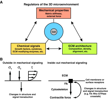
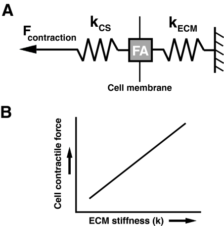
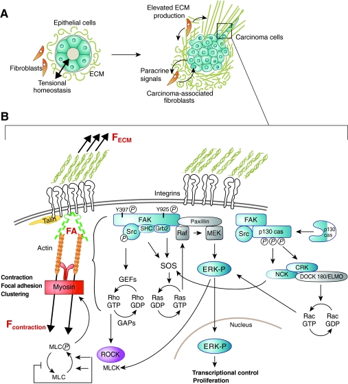
The notion that cell shape and spreading can regulate cell proliferation has evolved over several years, but only recently has this been linked to forces from within and upon the cell. This emerging area of mechanical signaling is proving to be wide-spread and important for all cell types. The microenvironment that surrounds cells provides a complex spectrum of different, simultaneously active, biochemical, structural and mechanical stimuli. In this milieu, cells probe the stiffness of their microenvironment by pulling on the extracellular matrix (ECM) and/or adjacent cells. This process is dependent on transcellular cell-ECM or cell-cell adhesions, as well as cell contractility mediated by Rho GTPases, to provide a functional linkage through which forces are transmitted through the cytoskeleton by intracellular force-generating proteins. This Commentary covers recent advances in the underlying mechanisms that control cell proliferation by mechanical signaling, with an emphasis on the role of 3D microenvironments and in vivo extracellular matrices. Moreover, as there is much recent interest in the tumor-stromal interaction, we will pay particular attention to exciting new data describing the role of mechanical signaling in the progression of breast cancer.


Figures

Fig. 1.
Fig. 1.
The mechanically active 3D microenvironment. (A) The primary factors of the 3D microenvironment that influence cell behavior. A dynamic dependence exists between all three factors, but here the focus is on mechanical signals to and from the ECM and the role these stimuli have in cell proliferation. (B) Outside–in (left) and inside–out (right) mechanical signals. During normal physiological function, cells and tissues in the body experience multi-axial loading that result from a complex superposition of external forces to produce stress in the cell. For example, tensile stress (σT), compressive stress (σC) and shear stress (τ; depicted as the result of fluid flow over the cell) are commonly applied to cells during normal physiological tissue function. Of course, for each of these stimuli there is an equal force that exists in the cell (not shown). Furthermore, during inside–out signaling, chemical energy is converted to mechanical energy in order to generate contractile forces within the cell and to impart stress on the ECM (not shown), which results in an elevated force balance at the FA that influences signal transduction within the cell.
Fig. 2.
Fig. 2.
Cell contraction force as a function of ECM stiffness. (A) Contractile force is transmitted within the actin cytoskeleton through the FA to the ECM (kCS and kECM represent the stiffness of the actin cytoskeleton and the ECM, respectively). (B) The magnitude of cell-generated contractile force is dependent upon the stiffness of the ECM (kECM). As the stiffness of the microenvironment increases, the magnitude of contractile force also increases in order to maintain tensional homeostasis. Of course, the cellular response to the mechanical properties of the microenvironment is further complicated because of the viscoelastic behavior of the ECM and within the cell itself (not shown; see Box 1). Although the time scale over which cells deform the matrix suggests that the process can be well described by understanding elastic behavior; the physiological implication of viscoelastic phenomena and the degree to which mechanical behavior at cellular scale is dominated by either elastic or viscous effects in a certain situation is not well understood – but is likely to provide additional insight into mechanotransduction.
Fig. 3.
Fig. 3.
Inside–out contractile force as a regulator of cell signaling. Stiffness of the extracellular matrix influences the magnitude of the contractile force of the cell that is transmitted to the ECM through integrins. (A) Increased ECM deposition by carcinoma-associated fibroblasts (CAFs) produces a stiff microenvironment for carcinoma cells. The dense ECM contains CAFs that signal to carcinoma cells, provides micro- and/or nano-structures that influence cell behavior, and has a stiffness to which the cells respond. (B) Elevated stiffness of the extracellular matrix results in elevated forces (F) at the cell–matrix interface, and promotes 3D-matrix adhesion formation and maturation that leads to the activation of highly dynamic signaling networks that regulate fundamental cell processes such as proliferation.

References

    1. Aijaz S., D'Atri F., Citi S., Balda M. S., Matter K. (2005). Binding of GEF-H1 to the tight junction-associated adaptor cingulin results in inhibition of Rho signaling and G1/S phase transition. Dev. Cell 8, 777-786 - PubMed
    1. Alcaraz J., Xu R., Mori H., Nelson C. M., Mroue R., Spencer V. A., Brownfield D., Radisky D. C., Bustamante C., Bissell M. J. (2008). Laminin and biomimetic extracellular elasticity enhance functional differentiation in mammary epithelia. EMBO J. 27, 2829-2838 - PMC - PubMed
    1. Allinen M., Beroukhim R., Cai L., Brennan C., Lahti-Domenici J., Huang H., Porter D., Hu M., Chin L., Richardson A., et al. (2004). Molecular characterization of the tumor microenvironment in breast cancer. Cancer Cell 6, 17-32 - PubMed
    1. Alowami S., Troup S., Al-Haddad S., Kirkpatrick I., Watson P. H. (2003). Mammographic density is related to stroma and stromal proteoglycan expression. Breast Cancer Res. 5, R129-R135 - PMC - PubMed
    1. Amatangelo M. D., Bassi D. E., Klein-Szanto A. J., Cukierman E. (2005). Stroma-derived three-dimensional matrices are necessary and sufficient to promote desmoplastic differentiation of normal fibroblasts. Am. J. Pathol. 167, 475-488 - PMC - PubMed

Publication types

Substances