. 2011 Mar;7(3):e1001318.
doi: 10.1371/journal.ppat.1001318. Epub 2011 Mar 17.

Dendritic cells and hepatocytes use distinct pathways to process protective antigen from plasmodium in vivo

Affiliations

Dendritic cells and hepatocytes use distinct pathways to process protective antigen from plasmodium in vivo

Ian A Cockburn et al. PLoS Pathog. 2011 Mar.

Abstract

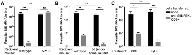
Malaria-protective CD8+ T cells specific for the circumsporozoite (CS) protein are primed by dendritic cells (DCs) after sporozoite injection by infected mosquitoes. The primed cells then eliminate parasite liver stages after recognizing the CS epitopes presented by hepatocytes. To define the in vivo processing of CS by DCs and hepatocytes, we generated parasites carrying a mutant CS protein containing the H-2K(b) epitope SIINFEKL, and evaluated the T cell response using transgenic and mutant mice. We determined that in both DCs and hepatocytes CS epitopes must reach the cytosol and use the TAP transporters to access the ER. Furthermore, we used endosomal mutant (3d) and cytochrome c treated mice to address the role of cross-presentation in the priming and effector phases of the T cell response. We determined that in DCs, CS is cross-presented via endosomes while, conversely, in hepatocytes protein must be secreted directly into the cytosol. This suggests that the main targets of protective CD8+ T cells are parasite proteins exported to the hepatocyte cytosol. Surprisingly, however, secretion of the CS protein into hepatocytes was not dependent upon parasite-export (Pexel/VTS) motifs in this protein. Together, these results indicate that the presentation of epitopes to CD8+ T cells follows distinct pathways in DCs when the immune response is induced and in hepatocytes during the effector phase.


Conflict of interest statement

The authors have declared that no competing interests exist.

Figures

Figure 1
Figure 1. Generation of P. berghei CS5M parasites.
A. Scheme of the strategy used for gene targeting of the replacement CS5M molecule. Location of primers used for PCR verification of recombination is given below (primer sequences given in Table S1). Restriction sites are K – KpnI; Se – SexAI; Bs – BsmF1; X – XhoI; S – SacI. B. Verification of clones – i. genomic DNA from cloned parasites was amplified with the primers CS1 and S8R (giving a 1526 bp product) to verify recombination at the 5′ end, and the primers CS4 and PB106 (giving a 1001 bp product) to verify recombination at the 3′ end, genomic DNA from P. berghei ANKA was used as a control. ii. To verify that the parasite population was clonal, genomic DNA was amplified within the CS sequence with the primers F205 and R904 to give a 699 bp product. The PCR product was then digested with SexA1, which cuts in the P. berghei CS5M product, but not the P. berghei ANKA product, to yield fragments of 510 and 186 bp. C. C57Bl/6 mice were immunized i.d. in the right ear with 5×10⁴ irradiated P. berghei ANKA or P. berghei CS5M parasites. 10 days later the SIINFEKL-specific immune response in the spleen, draining lymph nodes and liver (pooled) was determined by ELISPOT (mean ± SEM; n = 3, data from one of 3 similar experiments; **  =  P<0.01). D. C57Bl/6 mice received 2×10⁶ SIINFEKL-specific effector CD8+ T cells 3 hours prior to challenge with 5×10³ P. berghei ANKA or P. berghei CS5M sporozoites (grey bars); control mice did not receive effector cells (black bars). 40 hours later livers were taken and parasite rRNA concentration determined by real-time PCR (mean ± SEM; n = 4, data from one of 2 similar experiments, ns  =  not significant).
Figure 2
Figure 2. Antigen presentation by DCs is TAP dependent.
A. CFSE profiles of SIINFEKL specific transgenic cells after incubation with dLN DCs isolated from C57Bl/6 (wild type) or TAP-1 deficient animals 2 days after immunization with 5×10⁴ P. berghei CS5M sporozoites/ear. Data are based on pooled DCs from 6–8 mice per group; values at top left are the percent of cells that have divided. B. TAP1-/- and C57Bl/6 (wild type) mice received 2×10³ naïve SIINFEKL-specific CD8+ T cells prior to being fed on by 10–20 P. berghei CS5M infected mosquitoes. 10 days later the mice were sacrificed and the expansion of SIINFEKL-specific cells in the spleen and liver determined. Data are pooled from 2 similar experiments (mean ± SEM; n = 6).
Figure 3
Figure 3. Antigen presentation by DCs occurs via the endosome-to-cytosol pathway.
A. i. DCs were purified from the ear draining LN of C57Bl/6 (wild type) or Unc93B13d animals 2 days after immunization with 5×10⁴ P. berghei CS5M sporozoites/ear and incubated with CFSE labeled SIINFEKL specific trangenic cells. i Representative CFSE profiles of the transgenic cells 3 days after immunization values at top left are the percent of cells that have divided (mean ± SEM). ii. Mean number of cells that had divided at least one or twice in 3 independent experiments after incubation with DCs from wild type (black bars) or 3d mice (gray bars) (mean ± SEM; *  =  P<0.05; for each group in each experiment pooled DCs from 6–8 immunized animals were used). B. 3d (endosomal mutant) and C57Bl/6 mice received 2×10³ naïve SIINFEKL-specific cells prior to being fed on by 10–20 P. berghei CS5M infected mosquitoes. 10 days later the mice were sacrificed and the expansion of SIINFEKL-specific cells in the spleen and liver determined. Data are pooled from 2 similar experiments (mean ± SEM; n = 6; ***  =  P<0.001). C. C57Bl/6 mice received 2×10³ naïve SIINFEKL-specific cells prior to being fed on by 10–20 P. berghei CS5M infected mosquitoes. Treated mice received 15 mg of horse cyt c (Sigma) for 3 days starting on the day before immunization, control mice received vehicle alone (PBS). 10 days later the mice were sacrificed and the expansion of SIINFEKL-specific cells in the spleen and liver determined. Data are pooled from 2 similar experiments (mean ± SEM; n = 6).
Figure 4
Figure 4. Antigen presentation by DCs is inhibited by opsonization.
Mice received 5×10⁵ CFSE labeled SIINFEKL-specific cells one day prior to immunization i.d. with 5×10⁴ P. berghei CS5M parasites in the right ear that had either been treated for 20 minutes with 100 µg/ml of either anti-P. knowlesi CS (2G3), anti-P. berghei CS (3D11) or F(ab′)2 fragments prepared from the 3D11 antibody. Three days later the mice were sacrificed and ear draining lymph nodes taken. A. Representative CFSE profiles of the SIINFEKL-specific population, values are the mean % of cells that had proliferated ± SEM in one of 4 similar experiments. B. The size of the expansion of the transferred SIINFEKL-specific cells (mean ± SEM n = 3; representative of 4 similar experiments).
Figure 5
Figure 5. Antigen is directly presented to effector cells by hepatocytes.
A. C57Bl/6 or TAP1-/- mice received 2×10⁶ SIINFEKL-specific effector CD8+ T cells 3 hours prior to challenge with 5×10³ P. berghei CS5M sporozoites (grey bars); control mice did not receive effector cells (black bars). 40 hours later livers were taken and parasite rRNA concentration determined by real-time PCR (mean ± SEM; n = 4, data from one of 2 similar experiments). B. C57Bl/6 or 3d mice received 2×10⁶ SIINFEKL specific effector CD8+ T cells 3 hours prior to challenge with 5×10³ P. berghei CS5M sporozoites (grey bars); control mice did not receive effector cells (black bars). 40 hours later livers were taken and parasite rRNA concentration determined by real-time PCR (mean ± SEM; n = 5, data from one of 2 similar experiments). C. Cyt c or PBS treated C57Bl/6 mice received 2×10⁶ SIINFEKL specific effector CD8+ T cells 3 hours prior to challenge with 5×10³ P. berghei CS5M sporozoites (grey bars); control mice did not receive effector cells (black bars). Treated mice received 15 mg cyt c for 3 days starting the day before challenge. 40 hours after challenge livers were taken and parasite rRNA concentration determined by real-time PCR (mean ± SEM; n = 4).
Figure 6
Figure 6. Pexel/VTS motifs are not required for the presentation of CD8+ epitopes in the CS protein.
A. Fluorescence microscopy of Hepa1–6 cells 6 hours after infection with P. berghei CS5M and P. berghei CS5MΔP1–2. Parasites were visualized by staining with anti-Plasmodium HSP70 (red) and the localization of the CS protein determined by staining with the 3D11 mAb (green). B. % of parasites with CS visible in the host cell based on microscopy performed as in A, (mean ± SEM; data are based on 3 independent experiments per parasite strain with 50 parasites imaged per experiment). C. C57Bl/6 mice received 2×10⁶ SIINFEKL specific effector CD8+ T cells 3 hours prior to challenge with 5×10³ P. berghei CS5M or P. berghei CS5MΔP1–2 sporozoites (grey bars); control mice did not receive effector cells (black bars). 40 hours later livers were taken and parasite rRNA concentration determined by real-time PCR (mean ± SEM; n = 4, data from one of 2 similar experiments). D. C57Bl/6 mice received 5×10⁵ CFSE labeled naïve SIINFEKL-specific cells one day prior to immunization i.d. with 5×10⁴ P. berghei CS5M or P. berghei CS5MΔP1–2 parasites in the right ear. Three days later the mice were sacrificed and ear draining lymph nodes taken. Antigen presentation in vivo was inferred by determining the % of SIINFEKL-specific cells that had proliferated (mean ± SEM; n = 3, data representative of 2 similar experiments).

Similar articles

Cited by

References

    1. Nussenzweig RS, Vanderberg J, Most H, Orton C. Protective immunity produced by the injection of x-irradiated sporozoites of plasmodium berghei. Nature. 1967;216:160–162. - PubMed
    1. Romero P, Maryanski JL, Corradin G, Nussenzweig RS, Nussenzweig V, et al. Cloned cytotoxic T cells recognize an epitope in the circumsporozoite protein and protect against malaria. Nature. 1989;341:323–326. - PubMed
    1. Schofield L, Villaquiran J, Ferreira A, Schellekens H, Nussenzweig R, et al. Gamma interferon, CD8+ T cells and antibodies required for immunity to malaria sporozoites. Nature. 1987;330:664–666. - PubMed
    1. Chakravarty S, Cockburn IA, Kuk S, Overstreet MG, Sacci JB, et al. CD8+ T lymphocytes protective against malaria liver stages are primed in skin-draining lymph nodes. Nat Med. 2007;13:1035–1041. - PubMed
    1. Jobe O, Donofrio G, Sun G, Liepinsh D, Schwenk R, et al. Immunization with radiation-attenuated Plasmodium berghei sporozoites induces liver cCD8alpha+DC that activate CD8+T cells against liver-stage malaria. PLoS One. 2009;4:e5075. - PMC - PubMed

Publication types

MeSH terms