. 2011 Mar 18;6(3):e14762.
doi: 10.1371/journal.pone.0014762.

Serum IGF-1 affects skeletal acquisition in a temporal and compartment-specific manner

Affiliations

Serum IGF-1 affects skeletal acquisition in a temporal and compartment-specific manner

Hayden-William Courtland et al. PLoS One. .

Abstract

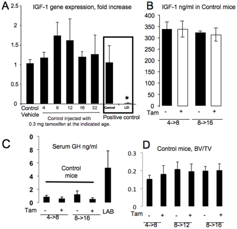
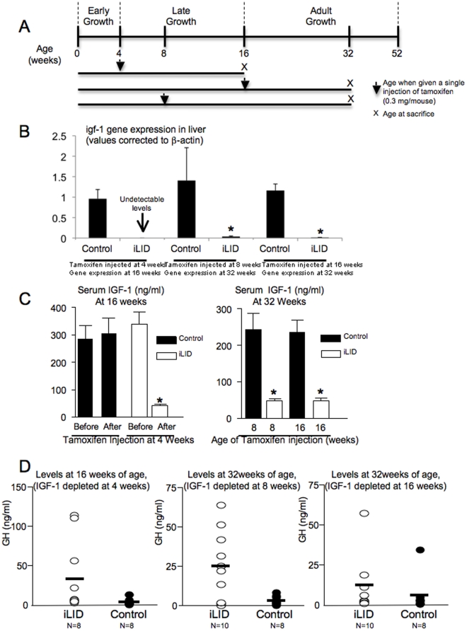
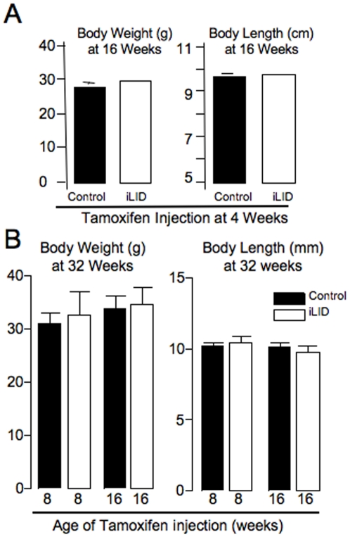
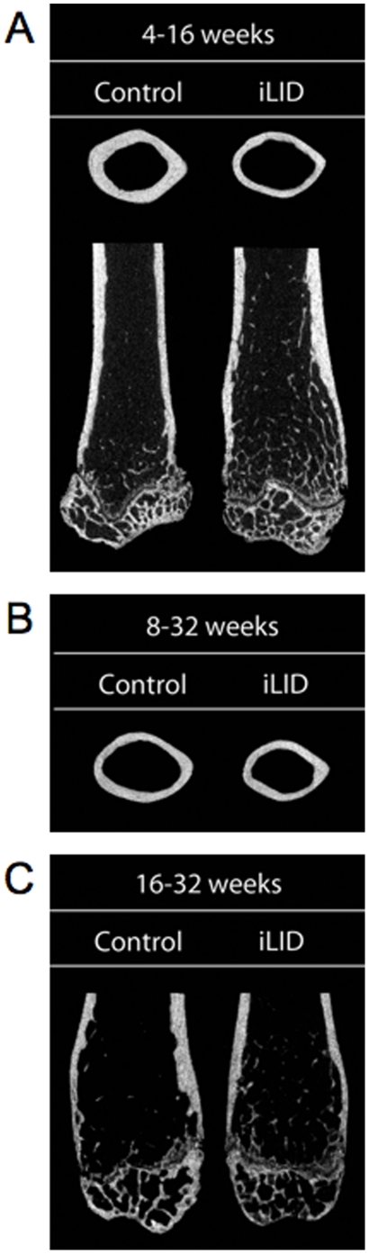
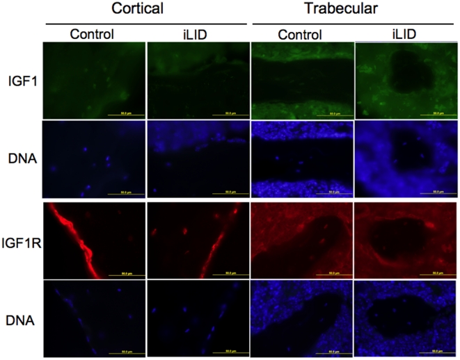
Insulin-like growth factor-1 (IGF-1) plays a critical role in the development of the growing skeleton by establishing both longitudinal and transverse bone accrual. IGF-1 has also been implicated in the maintenance of bone mass during late adulthood and aging, as decreases in serum IGF-1 levels appear to correlate with decreases in bone mineral density (BMD). Although informative, mouse models to date have been unable to separate the temporal effects of IGF-1 depletion on skeletal development. To address this problem, we performed a skeletal characterization of the inducible LID mouse (iLID), in which serum IGF-1 levels are depleted at selected ages. We found that depletion of serum IGF-1 in male iLID mice prior to adulthood (4 weeks) decreased trabecular bone architecture and significantly reduced transverse cortical bone properties (Ct.Ar, Ct.Th) by 16 weeks (adulthood). Likewise, depletion of serum IGF-1 in iLID males at 8 weeks of age, resulted in significantly reduced transverse cortical bone properties (Ct.Ar, Ct.Th) by 32 weeks (late adulthood), but had no effect on trabecular bone architecture. In contrast, depletion of serum IGF-1 after peak bone acquisition (at 16 weeks) resulted in enhancement of trabecular bone architecture, but no significant changes in cortical bone properties by 32 weeks as compared to controls. These results indicate that while serum IGF-1 is essential for bone accrual during the postnatal growth phase, depletion of IGF-1 after peak bone acquisition (16 weeks) is compartment-specific and does not have a detrimental effect on cortical bone mass in the older adult mouse.


Conflict of interest statement

Competing Interests: The authors have declared that no competing interests exist.

Figures

Figure 1
Figure 1. Validation of the iLID model.
(A) Liver expression of IGF-1 in control mice (mean ± SE) is not altered following 0.3 mg tamoxifen injection. Control mice at 4, 8, 12, 16 and 32 weeks of age were injected with 0.3 mg tamoxifen and livers were dissected four weeks later for gene expression analysis using real time PCR (n = 3 per group). Control and “constitutive” LID mice were used as positive control for the assay. Cycle number was corrected to β-actin and levels related to vehicle control, which was set as 1. (B) Mean serum IGF-1 levels in control mice (± SE) injected with 0.3 mg of tamoxifen (+) at 4 or 8 weeks were not altered compared to vehicle treated controls (−) at 8 or 16 weeks, respectively. (C) Mean serum GH levels in control mice (± SE) injected with 0.3 mg of tamoxifen (+) at 4 or 8 weeks were not altered compared to vehicle treated controls (−) at 8 or 16 weeks, respectively. Sera from triple knockout of LID/acid labile subunit KO/IGFBP-3KO (LAB) mice, which have high serum GH levels , served as a positive control. (D) BV/TV (bone volume/total volume) from micro-CT analysis of control mice injected with vehicle (−) or 0.3 mg tamoxifen (+) at 4, 8, and 12 weeks of age and then analyzed at 8, 12, and 16 weeks, respectively. Trabecular bone was analyzed at the femoral distal metaphysis (n = 6 per group). No significant differences were found between vehicle and 0.3 mg tamoxifen injected mice.
Figure 2
Figure 2. Experimental design, gene expression and serum levels for control and iLID mice.
A) Schematic representation of the experimental design. (B) igf-1 gene expression in liver in control and iLID mice injected with tamoxifen at different ages (C) Mean serum IGF-1 levels (± s.d) in control (n = 6) and iLID (n = 6 each) before (4 weeks) and after (16 weeks) injection of 0.3 mg of tamoxifen at 4 weeks. Serum IGF-1 levels in iLID mice are statistically different from control mice as expected (ANOVA, p<0.05). (D) Serum GH levels in control (n = 8) and iLID mic (n = 10) after depletion of serum IGF-1 by tamoxifen injection.
Figure 3
Figure 3. Body weight and length data for control and iLID mice.
(A) Mean body weight and body length (± SE) of control (n = 6) and iLID (n = 6) mice at 16 weeks after injection of 0.3 mg of tamoxifen at 4 weeks. (B) Body weights and body lengths (± s.d) for 32 week control and iLID after injection of tamoxifen at either 8 or 16 weeks of age.
Figure 4
Figure 4. Micro-CT images of control and iLID mice indicating differences in cortical and trabecular bone accrual.
(A) Mice injected with tamoxifen at 4 weeks and sacrificed at 16 weeks. (B) Mice injected with tamoxifen at 8 weeks and sacrificed at 32 weeks. (C) Mice injected with tamoxifen at 16 weeks and sacrificed at 32 weeks.
Figure 5
Figure 5. Representative immunofluroescene images of cortical and trabecular bone from 32 week control and iLID mice that were serum IGF-1 depleted at 16 weeks of age.
Antibody staining for IGF-1 and the IGF-1R showed no differences when comparing control and iLID mice.

Similar articles

Cited by

References

    1. Canalis E, Pash J, Varghese S. Skeletal growth factors. Crit Rev Eukaryot Gene Expr. 1993;3:155–166. - PubMed
    1. McCarthy TL, Centrella M, Canalis E. Regulatory effects of insulin-like growth factors I and II on bone collagen synthesis in rat calvarial cultures. Endocrinology. 1989;124:301–309. - PubMed
    1. Canalis E, Rydziel S, Delany AM, Varghese S, Jeffrey JJ. Insulin-like growth factors inhibit interstitial collagenase synthesis in bone cell cultures. Endocrinology. 1995;136:1348–1354. - PubMed
    1. Rydziel S, Delany AM, Canalis E. Insulin-like growth factor I inhibits the transcription of collagenase 3 in osteoblast cultures. J Cell Biochem. 1997;67:176–183. - PubMed
    1. Holly J. Physiology of the IGF system. Novartis Found Symp 262: 19–26; disussion. 2004:26–35. 265-268. - PubMed

Publication types

Substances