. 2011 Mar 18;6(3):e17830.
doi: 10.1371/journal.pone.0017830.

Histone demethylase JMJD2B functions as a co-factor of estrogen receptor in breast cancer proliferation and mammary gland development

Affiliations

Histone demethylase JMJD2B functions as a co-factor of estrogen receptor in breast cancer proliferation and mammary gland development

Masahito Kawazu et al. PLoS One. .

Erratum in

Abstract

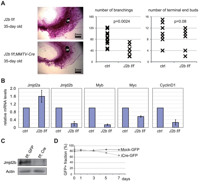
Estrogen is a key regulator of normal function of female reproductive system and plays a pivotal role in the development and progression of breast cancer. Here, we demonstrate that JMJD2B (also known as KDM4B) constitutes a key component of the estrogen signaling pathway. JMJD2B is expressed in a high proportion of human breast tumors, and that expression levels significantly correlate with estrogen receptor (ER) positivity. In addition, 17-beta-estradiol (E2) induces JMJD2B expression in an ERα dependent manner. JMJD2B interacts with ERα and components of the SWI/SNF-B chromatin remodeling complex. JMJD2B is recruited to ERα target sites, demethylates H3K9me3 and facilitates transcription of ER responsive genes including MYB, MYC and CCND1. As a consequence, knockdown of JMJD2B severely impairs estrogen-induced cell proliferation and the tumor formation capacity of breast cancer cells. Furthermore, Jmjd2b-deletion in mammary epithelial cells exhibits delayed mammary gland development in female mice. Taken together, these findings suggest an essential role for JMJD2B in the estrogen signaling, and identify JMJD2B as a potential therapeutic target in breast cancer.


Conflict of interest statement

Competing Interests: The authors have declared that no competing interests exist.

Figures

Figure 1
Figure 1. Elevated expression of JMJD2B in ER-positive breast cancers.
(A) JMJD2B mRNA expression in clinical breast cancer samples from 19 studies registered in the ONCOMINE database. Blue bars, JMJD2B mRNA in ER-negative samples; red bars, JMJD2B mRNA in ER-positive samples. Data were analyzed using ONCOMINE algorithms. mRNA expression levels are represented by normalized units. The line within each colored box represents the median value for each group, and the upper and lower edges of each box indicate the 75th and 25th percentiles, respectively. Study details and statistical values are described in Table S1. (B) JMJD2B protein levels are higher in ER-positive breast cancer cell lines. Extracts of ER-positive and ER-negative breast cancer cell lines were analyzed by western blot to detect JMJD2 family members. Actin, loading control. (C) JMJD2B protein expression is induced by E2 stimulation. T-47D cells (ER-positive) and MDA-MB468 cells (ER-negative) were cultured in steroid-depleted serum for 4 days and stimulated with or without E2 for 24 hr. Cell extracts were analyzed by western blot. Relative intensity of the JMJD2A and JMJD2B bands are shown. The values are normalized to the bands of corresponding actin. (D) E2 stimulates JMJD2B expression. T-47D cells were cultured in steroid-depleted serum for 4 days followed by E2 stimulation for 6 hr. JMJD2A, JMJD2B and JMJD2C transcript levels are shown relative to levels in untreated controls (set to a value of 1). Expression levels were normalized to ACTB expression levels. Data represent mean ± s.d. of triplicates. ** p<0.01. Full-length blots are presented in Figure S7.
Figure 2
Figure 2. JMJD2B positively regulates the proliferation of ER-positive breast cancer cells.
(A) JMJD2B knockdown impairs proliferation. T-47D cells were transfected with either control siRNA or siRNA against JMJD2A or JMJD2B, cultured for 72 hr, pulsed for 1 hr with BrdU, stained with APC-conjugated anti-BrdU antibody and 7-AAD, and analyzed by flow cytometry. Results are representative of four independent trials. (B) JMJD2B knockdown impairs colony formation. Single cell suspensions of ZR-75-1 cells expressing control shRNA or shRNA against JMJD2B were seeded in soft agar. After 14 days, colonies were stained with crystal violet and microscopic fields were photographed (left). The number of colonies/cm2 was determined (right). Data represent the colony density of three wells (mean ± s.d). **p<0.01. Representative data of three independent trials. See also Figure S2. (C) JMJD2B knockdown impairs tumor formation in xenograft model. Upper left: tumors (indicated by arrows) in a representative NIH-III mouse injected with control ZR-75-1 cells on the left flank and JMJD2B-depleted ZR-75-1 cells on the right flank. Upper right: dissected tumors from nude mice. Lower: mass of tumors is shown. **p<0.01.
Figure 3
Figure 3. JMJD2B interacts with ERα and SWI/SNF-B complex.
(A) JMJD2B co-immunoprecipitates with ERα. 293T cells were co-transfected with expression plasmids for FLAG-JMJD2B and either MYC-ERα or empty vector. Cell lysate and α-MYC immunoprecipitates were analyzed by western blot with α-FLAG antibody. (B) Structure of JMJD2B deletion mutants. Pro rich, proline rich region. LRPLL, LRPLL motif. PHDx2, double PHD domain. TUDORx2, double TUDOR domain. Western blot analysis of mutants is shown in Figure S3A. (C) The JmjC domain is sufficient for co-immunoprecipitation with ERα. 293T cells were co-transfected with MYC-ERα and individual JMJD2B deletion mutant expression vectors (refer to B). Cell lysates and α-FLAG immunoprecipitates were analyzed by western blot with antibodies to the indicated proteins. Relative intensity of the bands of the mutants is shown, normalized to the bands of corresponding input. Arrowheads, ERα bands; *, α-FLAG antibody. (D) Association of JMJD2 proteins with ERα, SWI/SNF complex, or SWI/SNF-B complex. (E) Association of JMJD2B with ERα or SWI/SNF-B complex in the absence or presence of estrogen simulation. (D and E) Nuclear lysates of T-47D cells were immunoprecipitated with control IgG or antibodies to the indicated proteins. Input lysate and immunoprecipitated samples were immunoblotted using antibodies to the indicated proteins. See also Figure S3. Full-length blots are in Figure S7.
Figure 4
Figure 4. JMJD2B mediates induction of ER target genes and estrogen-dependent proliferation of breast cancer cells.
(A) Effect of JMJD2A or JMJD2B knockdown on induction of ER target genes. T-47D cells and MCF-7 cells were transfected with either control siRNA, JMJD2A siRNA or JMJD2B siRNA, cultured in steroid-free medium for 72 hr, and stimulated with or without E2 for 4 hr. (B) JMJD2B knockdown impairs ER target gene induction. T-47D cells were transfected with either control siRNA or JMJD2B siRNA, cultured in steroid-free medium for 72 hr, and stimulated with or without E2 for 4 hr. (A and B) mRNA levels of JMJD2B or the indicated ER target genes were measured by real-time RT-PCR. Results represent mRNA levels normalized to the level of ACTB mRNA (mean ± s.d. of triplicates). **p<0.01. (C) Venn diagram representation of the differentially expressed genes. The area of each circle reflects the number of genes contained in the respective categories. The heat map is in Figure S4C. The lists of the genes are in Tables S2, S3, S4, S5. (D) JMJD2B knockdown impairs cellular response to estrogen. T-47D cells or MCF-7 cells transfected with control siRNA or JMJD2B siRNA were cultured in steroid-free medium for 48 hr, stimulated for 24 hr with E2, labeled for 1 hr with BrdU, and stained with anti-BrdU antibody and 7-AAD. The fraction of BrdU-positive cells was determined by flow cytometry. A representative result from three independent experiments.
Figure 5
Figure 5. JMJD2B is recruited to the ER binding site; H3K9me3 is demethylated at the ER binding site.
(A) Chromatin immunoprecipitation (ChIP) assay of ERα and JMJD2B at indicated gene loci. T-47D cells were steroid-depleted for 96 hr and treated with E2 for 45 min or 4 hr. mRNA levels of JMJD2B and the indicated ER target genes in the corresponding samples are in Figure S5B. (B) ChIP assay of H3K9me3 at indicated gene loci. T-47D cells expressing control shRNA or shRNA against JMJD2B were steroid-depleted for 96 hr and treated with E2 for 4 hr. Results were normalized to input values, and expressed relative to untreated controls (set at 1). (C) ChIP assay of RNAPII in MYB gene. T-47D cells expressing control shRNA or JMJD2B shRNA were treated as in (B). Results were normalized to input values, and expressed relative to untreated controls (set at 1). (D) Presence of ERα and H3K9me3 within the MYB locus in T-47D cells. A region previously shown to be devoid of ERα was chosen as a negative control site (ER-neg) . Cells were treated as in (B). H3K9me3 ChIP signals were normalized to input chromatin and expressed relative to ChIP signals of the negative control site (set at 1). X-axis represents distance from TSS. (A–C) ChIP samples were quantified by real-time PCR. Data represent mean ± s.d. of triplicates. Representative data from three independent trials. **p<0.01; *p<0.05. ER binding sites assessed in this study are shown in Figure S5.
Figure 6
Figure 6. Conditional deletion of Jmjd2b in mammary epithelium results in defective mammary gland development.
(A) Defective development of Jmjd2b-deficient mammary gland. Carmine-stained whole-mount preparations of mammary glands from 5-week old Jmjd2b flox/flox;MMTV-Cre and Jmjd2b flox/flox mice (Ctrl∶control). Representative littermates are shown. LN, lymph node. The numbers of branching and terminal end buds within a fat pad were determined (Jmjd2b flox/flox n = 11; MMTV-Cre;Jmjd2b flox/flox: n = 6). (B) Impaired gene expression in Jmjd2b flox/flox; MMTV-Cre MECs. Lin MECs were isolated from Jmjd2b flox/flox;MMTV-Cre and Jmjd2b flox/flox mice. mRNA levels of Jmjd2b, Myb, CyclinD1 and c-myc were examined by real-time RT-PCR. Results represent mRNA levels normalized to the level of Actb mRNA (mean ± s.d. of five independent littermate pairs). (C) Western blot showing the absence of Jmjd2b protein in Cre-infected Jmjd2b flox/flox MECs. Full-length blots are in Figure S7 (D) Impaired proliferation of Jmjd2b-deficient MECs. Lin MECs were isolated from Jmjd2b flox/flox mice, infected with Mock-GFP or Cre-IRES-GFP (iCre-GFP) retrovirus, and cultured in vitro. Fractions of GFP-positive cells were determined at the indicated time points by FACS analysis. A representative result from three independent experiments is shown.

Similar articles

Cited by

References

    1. Jordan VC, O'Malley BW. Selective estrogen-receptor modulators and antihormonal resistance in breast cancer. J Clin Oncol. 2007;25:5815–5824. - PubMed
    1. Robinson-Rechavi M, Escriva Garcia H, Laudet V. The nuclear receptor superfamily. J Cell Sci. 2003;116:585–586. - PubMed
    1. Brzozowski AM, Pike AC, Dauter Z, Hubbard RE, Bonn T, et al. Molecular basis of agonism and antagonism in the oestrogen receptor. Nature. 1997;389:753–758. - PubMed
    1. Rosenfeld MG, Lunyak VV, Glass CK. Sensors and signals: a coactivator/corepressor/epigenetic code for integrating signal-dependent programs of transcriptional response. Genes Dev. 2006;20:1405–1428. - PubMed
    1. Lonard DM, Lanz RB, O'Malley BW. Nuclear receptor coregulators and human disease. Endocr Rev. 2007;28:575–587. - PubMed

Publication types

MeSH terms

Substances