Generation and evaluation of an IPTG-regulated version of Vav-gene promoter for mouse transgenesis
- PMID: 21445314
- PMCID: PMC3061885
- DOI: 10.1371/journal.pone.0018051
Generation and evaluation of an IPTG-regulated version of Vav-gene promoter for mouse transgenesis
Abstract
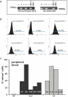
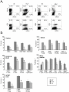
Different bacteria-derived systems for regulatable gene expression have been developed for the use in mammalian cells and some were also successfully adopted for in vivo use in vertebrate model organisms. However, certain limitations apply to most of these systems, including leakiness of transgene expression, inefficient transgene silencing or activation, as well as limited tissue accessibility of transgene-inducers or their unfavourable pharmacokinetics. In this study, we evaluated the suitability of the lac-operon/lac-repressor (lacO/lacI) system for the regulation of the well-established Vav-gene promoter that allows inducible transgene expression in different haematopoietic lineages in mice. Using the fluorescence marker protein Venus as a reporter, we observed that the lacO/lacI system could be amended to modulate transgene-expression in haematopoietic cells. However, reporter expression was not uniform and the lacO elements introduced into the Vav-gene promoter only conferred limited repression and reversion of lacI-mediated gene silencing after administration of IPTG. Although further optimization of the system is required, the lacO-modified version of the Vav-gene promoter may be adopted as a tool where low basal gene-expression and limited transient induction of protein expression are desired, e.g. for the activation of oncogenes or transgenes that act in a dominant-negative manner.
Conflict of interest statement
Figures


Similar articles
-
Regulation of endothelial-specific transgene expression by the LacI repressor protein in vivo.PLoS One. 2014 Apr 22;9(4):e95980. doi: 10.1371/journal.pone.0095980. eCollection 2014. PLoS One. 2014. PMID: 24755679 Free PMC article.
-
An engineered PGK promoter and lac operator-repressor system for the regulation of gene expression in mammalian cells.Gene. 1993 Aug 25;130(2):233-9. doi: 10.1016/0378-1119(93)90424-2. Gene. 1993. PMID: 8359690
-
Effect of isopropyl-beta-D-thiogalactopyranosid induction of the lac operon on the specificity of spontaneous and doxorubicin-induced mutations in Escherichia coli.Environ Mol Mutagen. 1995;26(1):16-25. doi: 10.1002/em.2850260104. Environ Mol Mutagen. 1995. PMID: 7641704
-
Regulation of the Escherichia coli lac operon expressed in human cells.Biochim Biophys Acta. 1992 Feb 28;1130(1):68-74. doi: 10.1016/0167-4781(92)90463-a. Biochim Biophys Acta. 1992. PMID: 1311956
-
Establishment of a low-dosage-IPTG inducible expression system construction method in Escherichia coli.J Basic Microbiol. 2018 Sep;58(9):806-810. doi: 10.1002/jobm.201800160. Epub 2018 Jul 2. J Basic Microbiol. 2018. PMID: 29962051
Cited by
-
Knockdown of the antiapoptotic Bcl-2 family member A1/Bfl-1 protects mice from anaphylaxis.J Immunol. 2015 Feb 1;194(3):1316-22. doi: 10.4049/jimmunol.1400637. Epub 2014 Dec 29. J Immunol. 2015. PMID: 25548219 Free PMC article.
-
Tracking of Engineered Bacteria In Vivo Using Nonstandard Amino Acid Incorporation.ACS Synth Biol. 2018 Jun 15;7(6):1640-1650. doi: 10.1021/acssynbio.8b00135. Epub 2018 Jun 6. ACS Synth Biol. 2018. PMID: 29791796 Free PMC article.
-
DNA-binding of the Tet-transactivator curtails antigen-induced lymphocyte activation in mice.Nat Commun. 2017 Oct 18;8(1):1028. doi: 10.1038/s41467-017-01022-4. Nat Commun. 2017. PMID: 29044097 Free PMC article.
-
The REMOTE-control system: a system for reversible and tunable control of endogenous gene expression in mice.Nucleic Acids Res. 2017 Dec 1;45(21):12256-12269. doi: 10.1093/nar/gkx829. Nucleic Acids Res. 2017. PMID: 28981717 Free PMC article.
-
Regulation of endothelial-specific transgene expression by the LacI repressor protein in vivo.PLoS One. 2014 Apr 22;9(4):e95980. doi: 10.1371/journal.pone.0095980. eCollection 2014. PLoS One. 2014. PMID: 24755679 Free PMC article.
References
-
- Adams JM, Harris AW, Strasser A, Ogilvy S, Cory S. Transgenic models of lymphoid neoplasia and development of a pan-hematopoietic vector. Oncogene. 1999;18:5268–5277. - PubMed
-
- Indra AK, Warot X, Brocard J, Bornert JM, Xiao JH, et al. Temporally-controlled site-specific mutagenesis in the basal layer of the epidermis: comparison of the recombinase activity of the tamoxifen-inducible Cre-ER(T) and Cre-ER(T2) recombinases. Nucleic Acids Res. 1999;27:4324–4327. - PMC - PubMed
-
- Gama Sosa MA, De Gasperi R, Elder GA. Animal transgenesis: an overview. Brain Struct Funct. 2010;214:91–109. - PubMed
-
- Corbel SY, Rossi FM. Latest developments and in vivo use of the Tet system: ex vivo and in vivo delivery of tetracycline-regulated genes. Curr Opin Biotechnol. 2002;13:448–452. - PubMed
Publication types
MeSH terms
Substances
Grants and funding
LinkOut - more resources
Full Text Sources
Molecular Biology Databases
Miscellaneous

