. 2011 Mar 21;6(3):e17550.
doi: 10.1371/journal.pone.0017550.

Location of intra- and extracellular M. tuberculosis populations in lungs of mice and guinea pigs during disease progression and after drug treatment

Affiliations

Location of intra- and extracellular M. tuberculosis populations in lungs of mice and guinea pigs during disease progression and after drug treatment

Donald R Hoff et al. PLoS One. .

Abstract

The lengthy treatment regimen for tuberculosis is necessary to eradicate a small sub-population of M. tuberculosis that persists in certain host locations under drug pressure. Limited information is available on persisting bacilli and their location within the lung during disease progression and after drug treatment. Here we provide a comprehensive histopathological and microscopic evaluation to elucidate the location of bacterial populations in animal models for TB drug development.To detect bacilli in tissues, a new combination staining method was optimized using auramine O and rhodamine B for staining acid-fast bacilli, hematoxylin QS for staining tissue and DAPI for staining nuclei. Bacillary location was studied in three animal models used in-house for TB drug evaluations: C57BL/6 mice, immunocompromised GKO mice and guinea pigs. In both mouse models, the bacilli were found primarily intracellularly in inflammatory lesions at most stages of disease, except for late stage GKO mice, which showed significant necrosis and extracellular bacilli after 25 days of infection. This is also the time when hypoxia was initially visualized in GKO mice by 2-piminidazole. In guinea pigs, the majority of bacteria in lungs are extracellular organisms in necrotic lesions and only few, if any, were ever visualized in inflammatory lesions. Following drug treatment in mice a homogenous bacillary reduction across lung granulomas was observed, whereas in guinea pigs the remaining extracellular bacilli persisted in lesions with residual necrosis. In summary, differences in pathogenesis between animal models infected with M. tuberculosis result in various granulomatous lesion types, which affect the location, environment and state of bacilli. The majority of M. tuberculosis bacilli in an advanced disease state were found to be extracellular in necrotic lesions with an acellular rim of residual necrosis. Drug development should be designed to target this bacillary population and should evaluate drug regimens in the appropriate animal models.


Conflict of interest statement

Competing Interests: The authors have declared that no competing interests exist.

Figures

Figure 1
Figure 1. Numbers of viable M. tuberculosis organisms in lungs of infected IFN-γ knock-out and C57BL/6 mice after drug treatment.
Mice were infected with M. tuberculosis strain Erdman and treated with drugs starting 18 to 21 days after aerosol infection; with isoniazid (INH), rifampin (RIF), ethambutol (EMB), gatifloxacin (Gati) or moxifloxacin (Moxi). Sacrifice times were at 2, 5 and 7 days of drug treatment. Data points represent mean log10 viable bacilli +/− standard error present in whole lung homogenates.
Figure 2
Figure 2. Intra-and extracellular M. tuberculosis bacilli in lungs throughout infection from M. tuberculosis infected GKO mice and after 9 days of INH drug treatment.
(AR) auramine-rhodamine, hematoxylin QS and DAPI; (H&E) hematoxylin and eosin. (A) An inflammatory lesion from lungs of an M. tuberculosis infected GKO mouse 18 days after aerosol infection. The inflammatory lesion shows a mix of macrophages and granulocytes arranged in an unorganized manner (H&E, 100× magnification). (B) Fluorescent image of an inflammatory lesion from lungs of an M. tuberculosis infected GKO mouse 18 days after aerosol infection. Intracellular red fluorescent AR+ stained bacilli were predominantly found uniformly distributed throughout inflammatory lesions at this time (AR, 200× magnification). (C) High magnification fluorescent image of the inflammatory lesion in figure 2B. The lesion shows intracellular AR+ bacilli located within various macrophage cells (AR, 400× magnification). (D) Cropped image taken from figure 2C (square) showing multiple AR+ stained bacilli within a single macrophage cell (AR, digital magnification). (E) A necrotic inflammatory lesion from lungs of an M. tuberculosis infected GKO mouse 28 days after aerosol infection. Multiple foci of intense basophilic staining are seen in alveolar spaces as they accumulate cellular necrotic debris (H&E staining, 40× magnification). (F) High magnification of alveolar spaces filled with cellular necrotic debris located within the inflammatory lesion depicted in figure 2E (square) (H&E staining, 400× magnification). (G) High magnification of a non-necrotic area within the inflammatory lesion seen in figure 2E (star) taken from a serial tissue section. Fluorescent image shows a number of intracellular AR+ stained bacilli residing within macrophages (AR, 1000× magnification). (H) High magnification of an alveolar space filled with necrotic debris located within the inflammatory lesion shown in figure 2E (circle) taken from a serial tissue section. Image shows a high number of extracellular AR+ stained bacilli residing among cellular necrotic debris situated in alveolar spaces (AR, 1000× magnification). (I) Fluorescent image of an inflammatory lesion from lungs of an M. tuberculosis infected GKO mouse 28 days after aerosol infection showing intracellular AR+ stained bacilli within multiple macrophages dispersed in non-granulomatous tissue (AR, 200× magnification). (J) AR+ bacilli within a remaining inflammatory lesion from lungs of an M. tuberculosis infected GKO mouse after 10 days of INH treatment (AR, 100× magnification). (K) Higher magnification of the inflammatory lesion seen in figure 2J. The lesion shows that the majority of remaining AR+ stained bacilli are intracellular within macrophages (AR, 200× magnification). (L) High magnification of the inflammatory lesion seen in figure 2K (square). This fluorescent image shows AR+ bacilli were predominantly found within macrophage cells comprising the few remaining inflammatory lesions (AR, 1000× magnification).
Figure 3
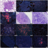
Figure 3. The progression of cellular necrosis that occurs within pulmonary inflammatory lesions during M. tuberculosis infection in GKO mice and the development of hypoxia as revealed by pimonidazole.
(AR) = auramine-rhodamine, hematoxylin QS and DAPI; (H&E) = hematoxylin and eosin. (A) An inflammatory lesion from the lungs of M. tuberculosis infected GKO mice 22 days after aerosol infection. The lesion shows a number of alveolar spaces beginning to accumulate granulocytes prior to the presence of cellular necrosis (H&E staining, 200× magnification). (B) High magnification of the area demarcated in figure 3A (circle) showing an alveolus completely occluded with inflammatory infiltrate (H&E staining, 1000× magnification). (C) Fluorescent image of an inflammatory lesion from the lungs of M. tuberculosis infected GKO mice 18 days post-aerosol infection showing a highly organized tubulin network with intact nuclei (Immunofluorescence for Tubulin (green) and MTB (red) and DAPI (blue). 1000× magnification). (D) Immunofluorescent image of an inflammatory lesion from the lungs of M. tuberculosis infected GKO mice 28 days post-aerosol TB (red) infection showing loss of tubulin (green) architecture and degenerated nuclei (blue) indicating loss of cell viability (1000× magnification). (E) Confocal microscopy of an inflammatory lesion from the lungs of M. tuberculosis infected GKO mice 18 days post-aerosol infection. The nuclei (blue) appear healthy with few numbers of AR+ bacilli (red). (AR staining combined with DAPI; 630× magnification with additional 2.5× digital zoom). (F) Confocal microscopy of an inflammatory lesion from the lungs of M. tuberculosis infected GKO mice 28 days post-aerosol infection. Degenerated fragments of inflammatory cell nuclei are within alveoli with a high number of extracellular AR+ bacilli (AR staining combined with DAPI; 630× magnification with additional 2.5× digital zoom). (G) Immunohistochemistry detecting hypoxia (brown) in a necrotic lung lesion from an M. tuberculosis infected GKO mouse 29 days after aerosol infection. The area of central necrosis (N) is surrounded by epithelioid macrophages near a major airway. (Hematoxylin counterstain, 40× magnification). (H) High magnification of the area demarcated within the inflammatory lesion shown in figure 3G (square). The center of alveoli, which is likely hypoxic, is filled with cellular debris and fails to stain due to the lack of viable cells (Hematoxylin counterstain, 400× magnification).
Figure 4
Figure 4. Intracellular M. tuberculosis bacilli in lungs from M. tuberculosis infected C57BL/6 mice, throughout infection and after 6 weeks of MXF drug treatment.
(AR) auramine-rhodamine, hematoxylin QS and DAPI; (H&E) hematoxylin and eosin. (A) An inflammatory lesion from the lungs of an M. tuberculosis infected C57BL/6 mouse 4 weeks after aerosol infection. The cellular architecture of lesions shows a field of lymphocytes (L) surrounding multiple macrophage aggregates (Φ) (H&E staining, 200× magnification). (B) An inflammatory lesion from lungs of an M. tuberculosis infected C57BL/6 mouse 4 weeks after aerosol infection. Intracellular AR+ bacilli were predominantly found within macrophage rich regions surrounded by lymphocytes (AR, 400× magnification). (C) High magnification of the inflammatory lesion shown in figure 4B (circle) showing intracellular AR+ stained bacilli within macrophages (AR, 1000× magnification). (D) An inflammatory lesion from the lungs of an M. tuberculosis infected C57BL/6 mouse 9 weeks after aerosol infection. The lesion shows a distinct rim of lymphocytes (L) surrounding a core of epithelioid macrophages (Φ). A layer of foamy macrophages (f) can be seen surrounding the lymphocyte rim (H&E staining, 200× magnification). (E & F) Fluorescent images of low (E) and high (F) magnifications of the inflammatory lesion depicted in figure 4D taken from a serial tissue section. The majority of AR+ bacilli at this time are found in epitheloid mΦs (Φ) located within the lymphocyte cuff (L), whereas a lower number of AR+ bacilli are located in foamy mΦs (f) located at the peripheral edges of inflammation (AR, 100× and 200× magnifications). (G) Inflammatory lesion from the lungs of a C57Bl/6 mouse infected with M. tuberculosis and treated with moxifloxacin for 6 weeks. Cellular architecture of lesions after 6 weeks of drug treatment is similar to untreated controls and consist of a mΦ core (Φ) surrounded by lymphocytes (L) and foamy mΦs (f) (AR, 200× magnification). (H) High magnification of the peripheral edges of the inflammatory lesion shown in figure 4G. A few remaining AR+ bacilli are found within foamy macrophages located outside of the lymphocyte cuff (AR, 1000× magnification).
Figure 5
Figure 5. Location of M. tuberculosis bacilli in lungs from M. tuberculosis infected guinea pigs, throughout infection and after 6 weeks of INH or TMC207 drug treatment.
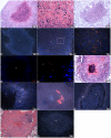
(AR) auramine-rhodamine, hematoxylin QS and DAPI; (H&E) hematoxylin and eosin. (A, B) Low (A) and high (B) magnifications of an early primary granuloma in the lungs of M. tuberculosis infected guinea pigs 4 weeks post-aerosol infection. Primary granulomas are distinguished from secondary lesions by the presence of necrosis (N) (H&E staining, 100× and 1000× magnifications). (C) A secondary lesion in the lungs of an M. tuberculosis infected guinea pig 4 weeks post-aerosol infection. Secondary lesions are distinguished from primary granulomas by the lack of central necrosis in the former (H&E staining, 100× magnification). (D, E) Low (D) and high (E) magnifications of a primary granuloma from the lungs of an M. tuberculosis infected guinea pig 4 weeks after aerosol infection. The majority of AR+ bacilli are extracellular within the necrotic core (c) (AR, 100× and 200× magnifications). (F) High magnification of area demarcated in figure 5E (square). Extracellular AR+ bacilli in the necrotic core of primary granulomas exist as single cells or are situated in clusters (AR, 1000× magnification). (G, H) Confocal micrographs of a necrotic granuloma showing an acellular necrotic core (c) with extracellular AR+ bacilli (red) (AR and DAPI, 200× and 630× magnifications). (I) A primary granuloma from the lungs of an M. tuberculosis infected guinea pig 10 weeks after aerosol infection showing advanced calcification and calcification of the necrotic core (c) and the acellular rim (R) surrounding the core (H&E staining, 100× magnification). (J) Fluorescent image of a primary lung granuloma from an M. tuberculosis infected guinea pig 10 weeks after aerosol infection. Extracellular AR+ bacilli are present within the necrotic core (c) and the acellular rim (R) surrounding the necrotic regions (AR, 200× magnification). (K) Cropped image from figure 5I (square) showing extracellular AR+ stained bacilli in the acellular rim. (L) Fluorescent image of a necrotic primary lung granuloma from an M. tuberculosis infected guinea pig treated for 6 weeks with INH. Extra-cellular AR+ bacilli are primarily within the core (c) of the partially calcified lytic necrosis, and to a lesser extent within the acellular, uncalcified rim (R) (AR, 200× and 400× magnifications). (M) A low magnification of the remnant of a primary lung granuloma in an M. tuberculosis infected guinea pig treated for 6 weeks with TMC207. The primary granuloma shows a caseous necrotic core (c) surrounded by inflammatory cells (H&E staining, 100× magnification). (N) Fluorescent image of the caseous necrotic core (c) in the primary lung granuloma shown in figure 5M taken from a serial tissue section. The few extra-cellular AR+ bacilli remaining after TMC207 treatment are primarily located within the central core of caseous necrosis (AR, 400× magnification).

References

    1. WHO annual report on global TB control–summary. Wkly Epidemiol Rec. 2003;78:122–128. - PubMed
    1. Trends in tuberculosis–United States, 2007. MMWR Morb Mortal Wkly Rep. 2008;57:281–285. - PubMed
    1. Gomez JE, McKinney JD. M. tuberculosis persistence, latency, and drug tolerance. Tuberculosis (Edinb) 2004;84:29–44. - PubMed
    1. Sacchettini JC, Rubin EJ, Freundlich JS. Drugs versus bugs: in pursuit of the persistent predator Mycobacterium tuberculosis. Nat Rev Microbiol. 2008;6:41–52. - PubMed
    1. Ehlers S. Lazy, dynamic or minimally recrudescent? On the elusive nature and location of the mycobacterium responsible for latent tuberculosis. Infection. 2009;37:87–95. - PubMed

Publication types

Substances

LinkOut - more resources