

doi: 10.1371/journal.pbio.1001037.
Epub 2011 Mar 22.
Regarding the amazing choreography of clathrin coats
Affiliations
- PMID: 21445329
- PMCID: PMC3062531
- DOI: 10.1371/journal.pbio.1001037
Item in Clipboard
Regarding the amazing choreography of clathrin coats
PLoS Biol.
2011 Mar.
No abstract available
Conflict of interest statement
The author has declared that no competing interests exist.
Figures
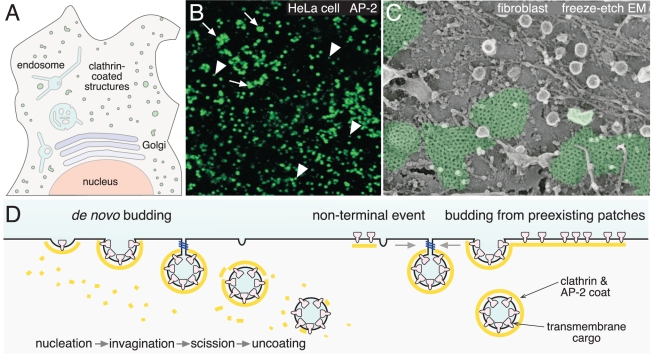
(A) A schematic bird's-eye view of a mammalian cell showing randomly
scattered clathrin-coated structures (green) positioned on the adherent cell
surface. (B) Confocal immunofluorescence image of the adherent surface of
HeLa cells stained with an antibody against the AP-2 adaptor protein showing
coexistence of diffraction-limited spots (arrowheads) and large clathrin
patches (arrows). (C) High-resolution electron micrograph of the adherent
surface of a cultured fibroblast (courtesy of John Heuser) showing areas of
flat clathrin lattice (pseudocolored in green). (D) Schematic depiction of
the process of clathrin-coated vesicle assembly at the various types of bud
site analyzed by Taylor et al. .
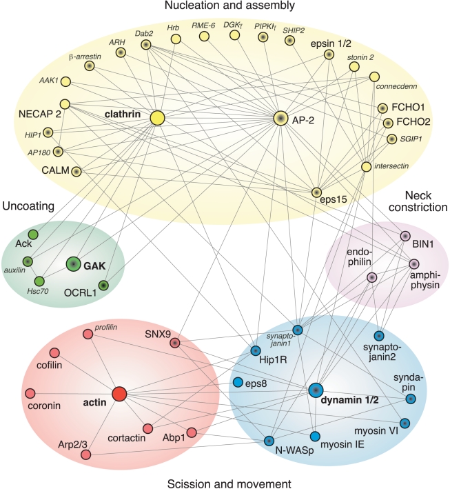
Hub-and-spoke depiction of a selected subset of the known proteins
participating in clathrin-mediated endocytosis. Established interactions are
indicated by the spokes. Modules are colored as in Taylor et al. and the
proteins they analyzed are shown in larger font. Note that not all of the
temporally defined modules are shown here. The symbols with black centers
indicate proteins that bind to phosphatidylinositol 4,5-bisphosphate, a
lipid marker of the plasma membrane. How can clathrin and AP-2 each bind to
so many partners (at once)? The functional clathrin molecule has at least 15
physically separate interaction surfaces while each AP-2 complex has over
ten.
References
-
- Schmid E. M, McMahon H. T. Integrating molecular and network biology to decode endocytosis. Nature. 2007;448:883–888. - PubMed
-
- Conner S. D, Schmid S. L. Regulated portals of entry into the cell. Nature. 2003;422:37–44. - PubMed
-
- Iwasa J. H. Animating the model figure. Trends Cell Biol. 2010;20:699–704. - PubMed
-
- Traub L. M. Tickets to ride: selecting cargo for clathrin-regulated internalization. Nat Rev Mol Cell Biol. 2009;10:583–596. - PubMed
MeSH terms
Substances
Grants and funding
LinkOut - more resources
Full Text Sources
