. 2011 Mar;69(3):445-54.
doi: 10.1002/ana.22337.

Demyelination causes synaptic alterations in hippocampi from multiple sclerosis patients

Affiliations

Demyelination causes synaptic alterations in hippocampi from multiple sclerosis patients

Ranjan Dutta et al. Ann Neurol. 2011 Mar.

Abstract

Objective: Multiple Sclerosis (MS) is an inflammatory demyelinating disease of the human central nervous system. Although the clinical impact of gray matter pathology in MS brains is unknown, 30 to 40% of MS patients demonstrate memory impairment. The molecular basis of this memory dysfunction has not yet been investigated in MS patients.

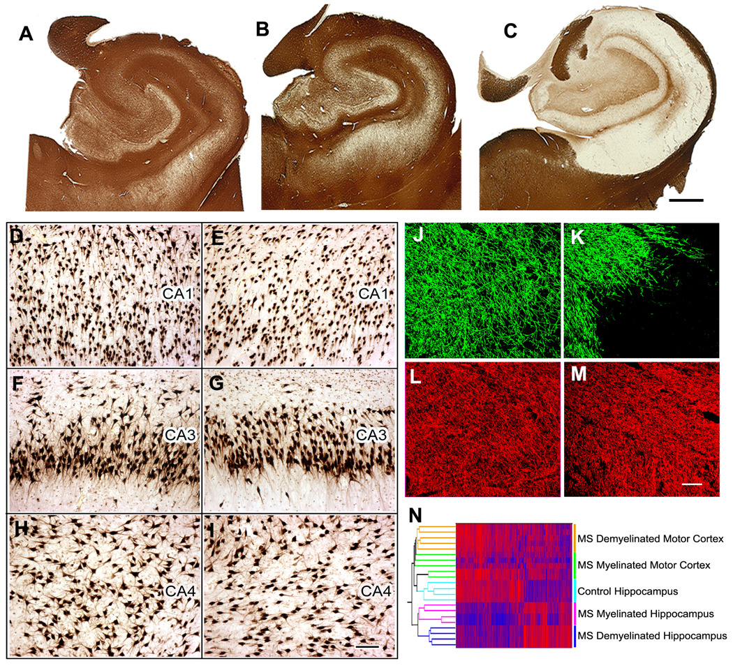
Methods: To investigate possible mechanisms of memory impairment in MS patients, we compared morphological and molecular changes in myelinated and demyelinated hippocampi from postmortem MS brains.

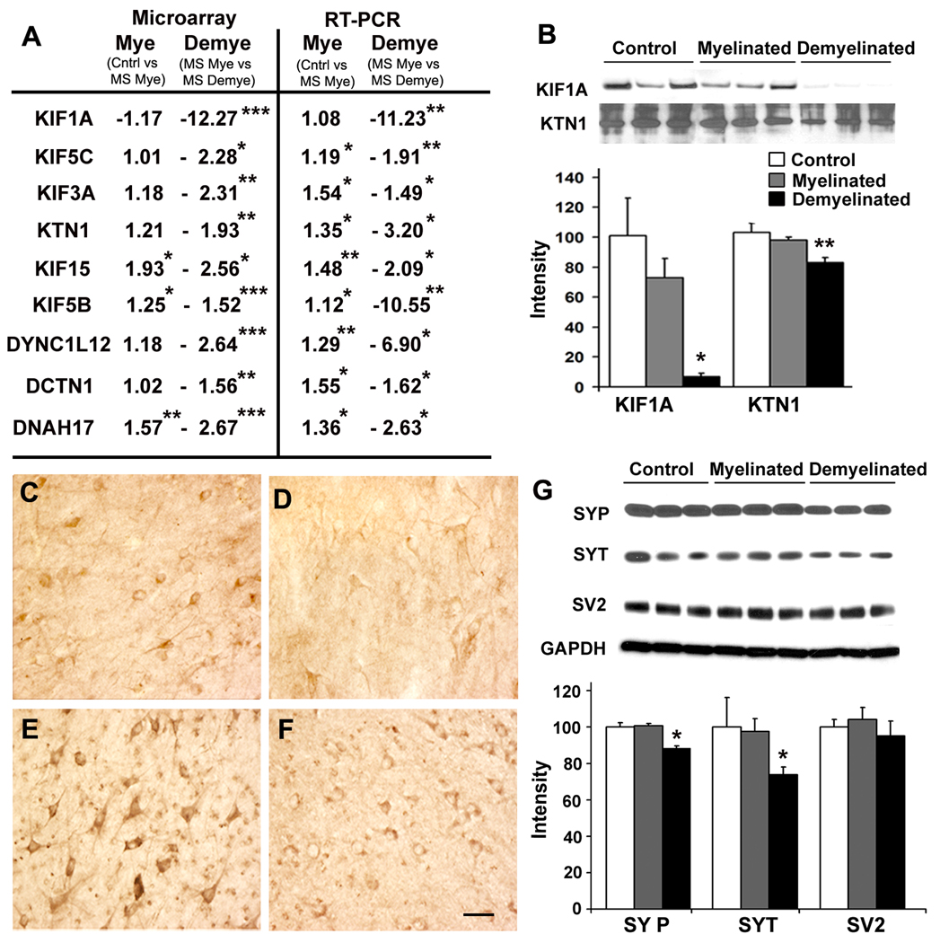
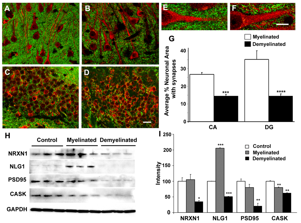
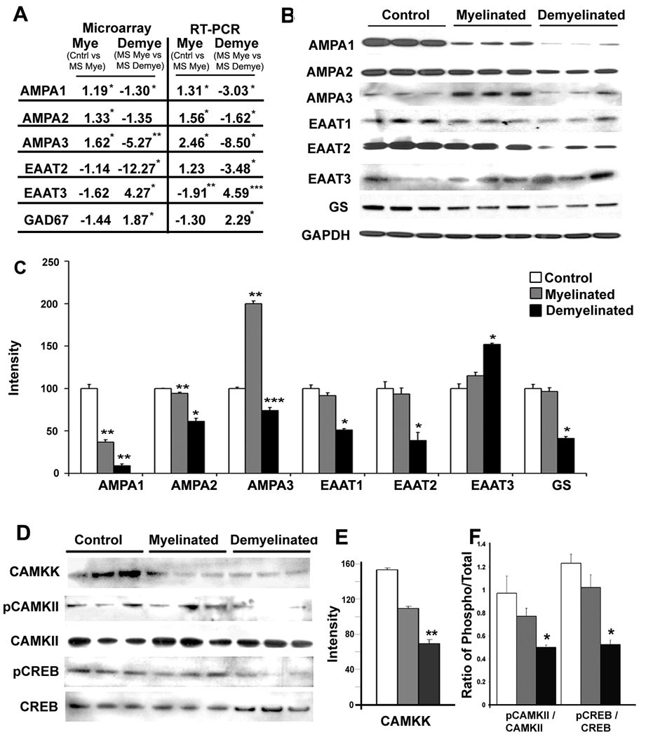
Results: Demyelinated hippocampi had minimal neuronal loss but significant decreases in synaptic density. Neuronal proteins essential for axonal transport, synaptic plasticity, glutamate neurotransmission, glutamate homeostasis, and memory/learning were significantly decreased in demyelinated hippocampi, but not in demyelinated motor cortices from MS brains.

Interpretation: Collectively, these data support hippocampal demyelination as a cause of synaptic alterations in MS patients and establish that the neuronal genes regulated by myelination reflect specific functions of neuronal subpopulations.


Figures

Figure 1
Figure 1
Hippocampal demyelination is common in multiple sclerosis (MS) brains and is associated with relative preservation of neurons and a distinct gene expression profile. (Panel A–C) Immunostaining for proteolipid protein (PLP) detected preservation of myelin in all control (A) and 10 of 22 MS hippocampi (B). Twelve of 22 MS hippocampi had significant or complete loss of myelin (C). (Panel D–I) Neuronal status in demyelinated MS hippocampus was determined by HuR immunohistochemistry. When compared to control hippocampi (Panels D, F, H), significant neuronal loss was not detected in CA1, CA3 or CA4 regions of demyelinated MS hippocampi (Panels E, G, I). (Panel J–M) Demyelinated hippocampal axons appear healthy. Double labeling immunofluorescence for myelin (myelin basic protein (MBP), green) and axons (SMI32, red) showed loss of myelin (K) with relative preservation of axons (M) in MS demyelinated hippocampus compared to control hippocampus (MBP, J; SMI32, I). (N) Demyelination alters hippocampal mRNA transcripts. Dual clustering of mRNA expression levels arranged samples into discrete clusters based upon myelin status (myelinated and demyelinated) and location (hippocampus vs motor cortex). High mRNA levels are indicated by red and blue denotes low expression levels. Scale bars: A–C: 2mm, D–I: 100µm, J–M:50µm
Figure 2
Figure 2
Hippocampal demyelination decreases proteins involved in axonal transport and enriched in synaptic vesicles. (Panels A–F) Both mRNA and protein transcripts of fast axonal transport genes were decreased in demyelinated MS hippocampal neurons. (A) mRNA encoding kinesin and dynein proteins were significantly decreased in microarray analysis (left), and verified by quantitative RT-PCR (right). mRNA levels of Kinesin 1A (KIF1A), Kinesin 5C (KIF5C), Kinesin 3A (KIF3A), Kinectin 1 (KTN1), Kinesin 15 (KIF15) Kinesin 5B (KIF5B), Dynein cytoplasmic light chain 12 (DYNC1L12), Dynactin 1 (DCTN1) and Dynein axonemal heavy polypeptide 17 (DNAH17) were significantly decreased in demyelinated compared to myelinated hippocampi. KIF1A and KTN1 were also reduced at the protein level (B). Immunohistochemistry shows presence of KIF1A and KTN1 in neurons in myelinated (Panels C, E) and demyelinated hippocampi (Panels D, F). (G) Synaptic vesicle proteins were decreased in demyelinated MS hippocampus. Protein levels of synaptophysin (SYP) and synaptotagmin (SYT) were significantly decreased in demyelinated MS hippocampus compared to myelinated MS hippocampus. Levels of synaptic vesicle glycoprotein-2, (SV2) were similar between control, myelinated MS and demyelinated MS hippocampi. GAPDH was used to verify equal loading of total protein. Scale Bars: C–F: 50µm, Error bars indicate S.E.M, * p<0.05, **p<0.005, ***p<0.0005
Figure 3
Figure 3
Hippocampal demyelination deceases pre-synaptic terminals and levels of proteins that help maintain synapses. (Panels A–F) Confocal images of MS hippocampus immunostained with antibodies specific to MAP2 (red) and synaptophysin (green) show punctuate pre-synaptic terminals (green) surrounding neurons in CA1 (Panels A, B) and dentate gyrus (Panels C, D) regions in MS myelinated (Panels A, C) and MS demyelinated (Panels B, D) hippocampus. Compared to myelinated MS hippocampi (E), pre-synaptic terminals were significantly decreased (G) in demyelinated MS hippocampi (F). (H) Levels of proteins essential for maintenance of synapses are decreased in demyelinated MS hippocampus. Neurexin 1 (NRXN1), neuroligin 1 (NLG1), post-synaptic density protein 95 (PSD95) and calmodulin-associated serine/threonin kinase (CASK) were significantly decreased in demyelinated MS hippocampi compared to myelinated MS hippocampi (I). GAPDH was used a loading control. Scale Bars: A–D: 20µm, E–F: 10µm, Error bars indicate S.E.M, * p<0.05, **p<0.005, ***p<0.0005, ****p<0.00005
Figure 4
Figure 4
Demyelination leads to decreased expression of glutamate receptors, glutamate transporters and key intermediates of memory/learning in MS hippocampus. (A–C) mRNA and protein levels of glutamate receptors and transporters were decreased in demyelinated MS hippocampus. (A) mRNAs encoding AMPA1, AMPA2, AMPA3, and EAAT2 were significantly decreased while mRNAs encoding EAAT3 and GAD67 were significantly increased in MS demyelinated hippocampi as measured by microarray analysis (left) and validated by quantitative RT-PCR (right). (B) Protein levels of AMPA receptors (AMPA1, AMPA2, AMPA3), glutamate transporters (EAAT1, EAAT2) and glutamine synthase (GS) were decreased in demyelinated MS hippocampi, while the neuronal glutamate transporter EAAT3 was significantly increased (C). (D–F) Key intermediates of memory/learning are decreased in demyelinated MS hippocampi. Phosphorylated CAMKII (E) and phosphorylated CREB (F), but not total CAMKII and total CREB, were significantly decreased in demyelinated MS hippocampi. Error bars indicate S.E.M., *p<0.05, ** P<0.005, ***p<0.0005

Comment in

References

    1. Trapp BD, Nave KA. Multiple sclerosis: an immune or neurodegenerative disorder? Annu Rev Neurosci. 2008;31:247–269. - PubMed
    1. Noseworthy JH. Progress in determining the causes and treatment of multiple sclerosis. Nature. 1999;399:A40–A47. - PubMed
    1. Chiaravalloti ND, DeLuca J. Cognitive impairment in multiple sclerosis. Lancet Neurol. 2008;7:1139–1151. - PubMed
    1. Bobholz JA, Rao SM. Cognitive dysfunction in multiple sclerosis: a review of recent developments. Curr Opin Neurol. 2003;16:283–288. - PubMed
    1. Rao SM, Leo GJ, Aubin-Faubert P. On the nature of memory disturbance in multiple sclerosis. J Clin Exp Neuropsychol. 1989;11:699–712. - PubMed

Publication types

MeSH terms

Substances