. 2011 Mar;69(3):540-52.
doi: 10.1002/ana.22275. Epub 2010 Dec 8.

DUX4, a candidate gene for facioscapulohumeral muscular dystrophy, causes p53-dependent myopathy in vivo

Affiliations

DUX4, a candidate gene for facioscapulohumeral muscular dystrophy, causes p53-dependent myopathy in vivo

Lindsay M Wallace et al. Ann Neurol. 2011 Mar.

Abstract

Objective: Facioscapulohumeral muscular dystrophy (FSHD) is associated with D4Z4 repeat contraction on human chromosome 4q35. This genetic lesion does not result in complete loss or mutation of any gene. Consequently, the pathogenic mechanisms underlying FSHD have been difficult to discern. In leading FSHD pathogenesis models, D4Z4 contractions are proposed to cause epigenetic changes, which ultimately increase expression of genes with myopathic potential. Although no gene has been conclusively linked to FSHD development, recent evidence supports a role for the D4Z4-encoded DUX4 gene in FSHD. In this study, our objective was to test the in vivo myopathic potential of DUX4.

Methods: We delivered DUX4 to zebrafish and mouse muscle by transposon-mediated transgenesis and adeno-associated viral vectors, respectively.

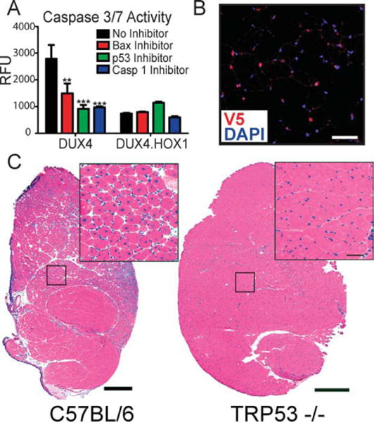
Results: Overexpression of DUX4, which encodes a transcription factor, caused abnormalities associated with muscular dystrophy in zebrafish and mice. This toxicity required DNA binding, because a DUX4 DNA binding domain mutant produced no abnormalities. Importantly, we found the myopathic effects of DUX4 were p53 dependent, as p53 inhibition mitigated DUX4 toxicity in vitro, and muscles from p53 null mice were resistant to DUX4-induced damage.

Interpretation: Our work demonstrates the myopathic potential of DUX4 in animal muscle. Considering previous studies showed DUX4 was elevated in FSHD patient muscles, our data support the hypothesis that DUX4 overexpression contributes to FSHD development. Moreover, we provide a p53-dependent mechanism for DUX4 toxicity that is consistent with previous studies showing p53 pathway activation in FSHD muscles. Our work justifies further investigation of DUX4 and the p53 pathway in FSHD pathogenesis.


Conflict of interest statement

Potential Conflicts of Interest

J.Y.: grants/grants pending, NIH. S.Q.H.: grants/grants pending, NIH.

Figures

FIGURE 1
FIGURE 1
DUX4 overexpression is detrimental to developing zebrafish muscle. (A) Tol2 zebrafish expression constructs contained striated muscle-specific MHCK7 promoter-driven DUX4 or hrGFP. ITR = inverted terminal repeat from Tol2 transposon; PA = SV40 polyA signal. (B) hrGFP epifluorescence showed MHCK7 activity in zebrafish muscle, which turned on 3 days postinjection. This lag in MHCK7 promoter expression allowed culling of abnormal embryos arising from nonspecific plasmid toxicity within the first 2 days postinjection. (C) MHCK7.DUX4 caused body malformation defects including short anterior-posterior (AP) axes, curved bodies, asymmetrically undeveloped pectoral fins (arrow indicates fin), or combinations of these morphologies. Some fish also showed cardiac hypertrophy (asterisk indicates heart) due to MHCK7-mediated DUX4 expression in the myocardium. (D) Hematoxylin and eosin staining of zebrafish body muscle shows MHCK7.DUX4 zebrafish had undefined somite boundaries, absent sarcomeric banding, and myofiber disorganization/degeneration. In contrast, MHCK7.hrGFP had no significant impact on gross body formation or somite/myofiber organization compared to normal zebrafish embryos. The only abnormal phenotypes seen in hrGFP fish were short AP axes, whereas undeveloped pectoral fins and abnormal body shapes were never present. Scale bars = 50mm. (E) Quantification of abnormal muscle phenotypes in zebrafish pictured in C–D. All DUX4-injected fish with abnormal body morphology also showed histological defects.
FIGURE 2
FIGURE 2
DUX4 toxicity requires DNA binding. (A) Structure of DUX4 adeno-associated virus (AAV) expression construct. White boxes indicate homeodomains (labeled 1 and 2). ITR = AAV inverted terminal repeats; CMV = cytomegalovirus promoter; PA = SV40 polyA signal. Alignment with a consensus homeodomain (Cons. Hox) identified 5 important residues required for DNA binding. *Indicates residues mutated to alanines in DUX4.HOX1 DNA binding mutant. (B) Western blot using extracts from transfected HEK293 cells showed DUX4.HOX1 protein was expressed at expected molecular weight (~50kDa) and consistently produced at higher levels than normal DUX4 in vitro. Lipo = HEK293 cells transfected with Lipofectamine-2000™ (Invitrogen, Carlsbad, CA) but no DNA. (C) Unlike DUX4, the DUX4.HOX1 mutant did not cause apoptosis in vitro, as indicated by lack of caspase-3/7 activation following transfection into HEK293 cells. ***Indicates significant differences from Lipofectamine controls, p < 0.0001 (analysis of variance; n = 3 independent experiments performed in triplicate). RFU = relative fluorescent units from caspase-3/7assay. (D) Electrophoretic mobility shift assay. Lanes 1 and 2 (DUX4 and DUX4 SS, respectively) show a shifted and super-shifted oligonucleotide (oligo) corresponding to the DUX4 binding site in the PITX1 promoter. Lane 3, DUX4.HOX1 has lower affinity for PITX1 promoter oligo. Mutation of the PITX1 binding site (mutPITX1) further reduces binding by DUX4, as previously reported, whereas no binding occurs between DUX4.HOX1 and the mutPITX1 site. Arrow indicates free PITX1 promoter probe; arrowhead indicates DUX4-bound PITX1 promoter sequence; asterisk indicates DUX4-PITX1 promoter complex supershifted with V5 antibody. SS = supershift.
FIGURE 3
FIGURE 3
DUX4 is toxic to adult mouse muscle in vivo. (A) hrGFP epifluorescence shows AAV6 transduction of adult mouse tibialis anterior (TA) 1 week postinjection. Hematoxylin and eosin (H&E) staining shows DUX4 caused massive myofiber degeneration and mononuclear cell infiltration that was not present in DUX4.HOX1 or hrGFP controls at high vector dose (3 × 10¹⁰ DRP). Scale bars = 500mm. (B) DUX4, but not DUX4.HOX1 or controls, significantly reduced TA muscle grip strength 1 and 2 weeks postinjection. n = 5 mice per group. ***p < 0.001; **p < 0.01; 2-way analysis of variance with Bonferroni post hoc test. H:F ratio indicates hindlimb (transduced) to forelimb (untransduced) grip strength ratios. (C) At lower doses (8 × 10⁸ DRP), DUX4 caused myofiber degeneration (by 1 week, shown here) that recapitulated the focal dystrophic lesions seen in facioscapulohumeral muscular dystrophy patients. Arrows point to degenerating myofibers, indicated by loss of acidophilic staining in H&E stains. Scale bars: left panel = 500mm; right panel = 50mm. (D) H&E staining revealed abundant centrally located nuclei and myofiber size variability only in DUX4-injected muscles. PI = indicates postinjection. Scale bar = 50mm. (E) Distribution of fiber diameter as a percentage of total fibers counted during sampling. DUX4 transduced muscles had more small-bore fibers compared to all controls, which is characteristic of regenerating dystrophic muscle. (F) DUX4-injected muscles had significantly higher percentages of centrally located nuclei (%C.N.) at both time points, which is another feature of dystrophic muscle, p < 0.001 (chi-square). All injections for panels C, D, and E delivered 8 × 10⁸ DRP of AAV6 vectors.
FIGURE 4
FIGURE 4
DUX4-transduced myofibers are caspase-3 positive. Top panels show DUX4+/caspase-3+ degenerating myofibers indicated by arrow, and shown in higher power in middle panels. Rabbit V5 antibody stain shows DUX4 was present in the nucleus but also had cytoplasmic localization in degenerating myofibers. In contrast, DUX4.HOX1 protein was exclusively nuclear. Some degenerating myofibers were caspase-3 negative but expressed DUX4 in the nucleus (caret) or cytoplasm (arrowhead). In contrast, several normal myofibers were DUX4+/caspase-3 negative (pound sign). Bottom panels, caspase-3 staining was absent in histologically normal muscle expressing DUX4.HOX1. The rabbit polyclonal caspase-3 primary antibody used here (Abnova; PAB0242) detects total caspase-3. Antibodies specifically recognizing cleaved caspase-3 showed similar staining patterns, as demonstrated in Supplementary Figure 3. DAPI (4′,6-diamidino-2-phenylindole) stains nuclear DNA. Scale bars = 50μm. H&E = hematoxylin and eosin.
FIGURE 5
FIGURE 5
DUX4 causes apoptosis through a p53-dependent mechanism. (A) DUX4-induced apoptosis is significantly reduced by Bax, p53, or caspase-1 inhibition, in vitro. **p < 0.01; ***p < 0.001 (analysis of variance). RFU = relative fluorescent units from caspase-3/7 assay. (B) V5 immunofluorescence and DAPI staining showed DUX4 expression in Trp53 −/− mouse myonuclei 2 weeks after injection. Scale bar = 50mm. (C) Trp53 −/− muscles are resistant to DUX4-induced degeneration, indicated by normal muscle histology in DUX4-transduced muscles, 2 weeks postinjection. In contrast, low-dose AAV6.DUX4 (8 × 10⁸ DRP) caused massive myofiber degeneration and subsequent regeneration 2 weeks postinjection. Scale bar = 500mm.

References

    1. Flanigan KM. Myology. 3. New York, NY: McGraw-Hill Medical Publishing Division; 2004. Facioscapulohumeral muscular dystrophy and scapuloperoneal disorders; pp. 1123–1133.
    1. Prevalence of rare diseases: bibliographic data. Orphanet. 2008 Available at: http://www.orpha.net/orphacom/cahiers/docs/GB/Prevalence_of_rare_disease... Accessed May 2010.
    1. Klinge L, Eagle M, Haggerty ID, et al. Severe phenotype in infantile facioscapulohumeral muscular dystrophy. Neuromuscul Disord. 2006;16:553–558. - PubMed
    1. van Deutekom JC, Wijmenga C, van Tienhoven EA, et al. FSHD associated DNA rearrangements are due to deletions of integral copies of a 3.2 kb tandem repeated unit. Hum Mol Genet. 1993;2:2037–2042. - PubMed
    1. Wijmenga C, Hewitt JE, Sandkuijl LA, et al. Chromosome 4q DNA rearrangements associated with facioscapulohumeral muscular dystrophy. Nat Genet. 1992;2:26–30. - PubMed

Publication types

MeSH terms

Substances