Review
. 2012 Jan;1821(1):137-51.
doi: 10.1016/j.bbalip.2011.03.005. Epub 2011 Apr 5.

Key enzymes of the retinoid (visual) cycle in vertebrate retina

Affiliations
Review

Key enzymes of the retinoid (visual) cycle in vertebrate retina

Philip D Kiser et al. Biochim Biophys Acta. 2012 Jan.

Abstract

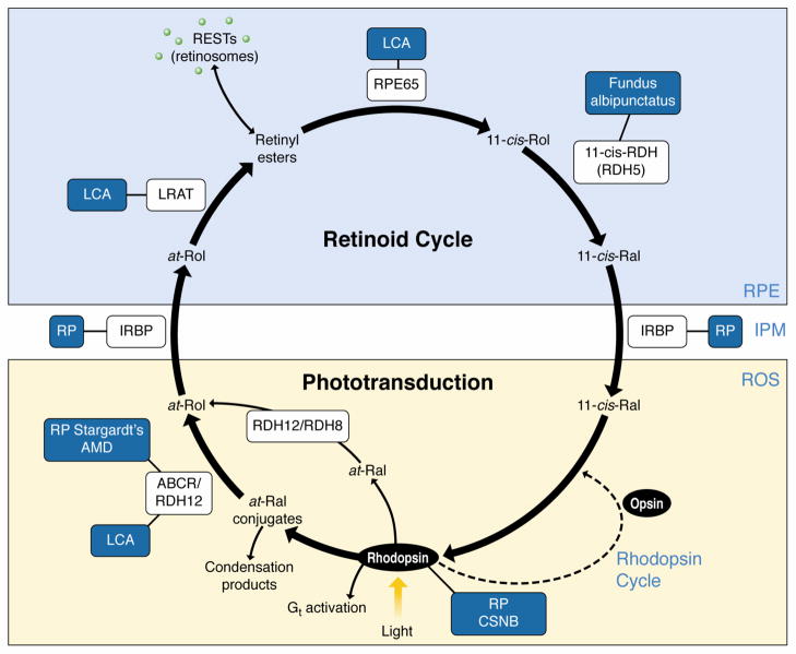
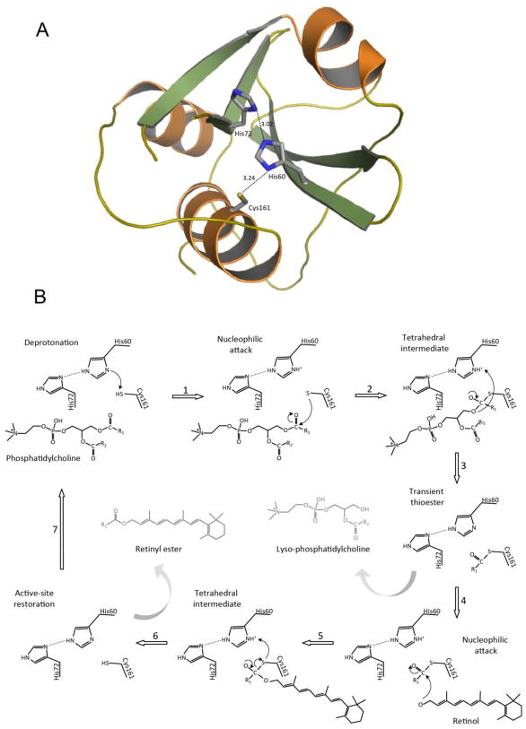
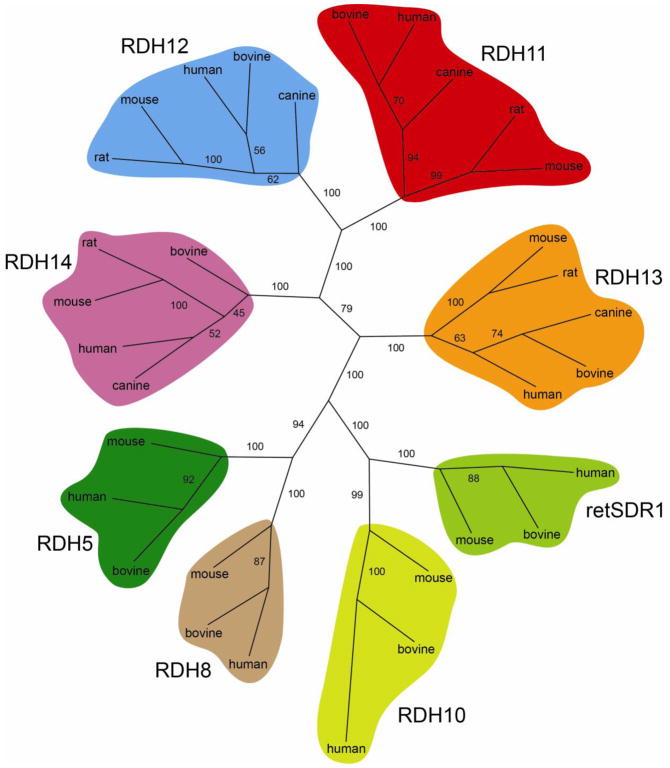
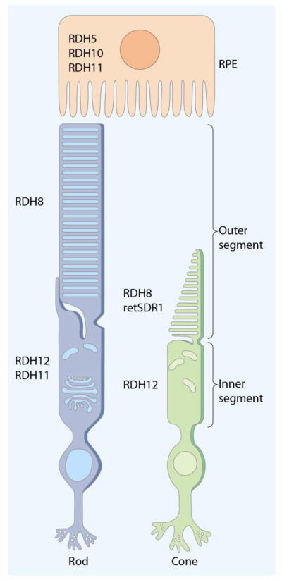
A major goal in vision research over the past few decades has been to understand the molecular details of retinoid processing within the retinoid (visual) cycle. This includes the consequences of side reactions that result from delayed all-trans-retinal clearance and condensation with phospholipids that characterize a variety of serious retinal diseases. Knowledge of the basic retinoid biochemistry involved in these diseases is essential for development of effective therapeutics. Photoisomerization of the 11-cis-retinal chromophore of rhodopsin triggers a complex set of metabolic transformations collectively termed phototransduction that ultimately lead to light perception. Continuity of vision depends on continuous conversion of all-trans-retinal back to the 11-cis-retinal isomer. This process takes place in a series of reactions known as the retinoid cycle, which occur in photoreceptor and RPE cells. All-trans-retinal, the initial substrate of this cycle, is a chemically reactive aldehyde that can form toxic conjugates with proteins and lipids. Therefore, much experimental effort has been devoted to elucidate molecular mechanisms of the retinoid cycle and all-trans-retinal-mediated retinal degeneration, resulting in delineation of many key steps involved in regenerating 11-cis-retinal. Three particularly important reactions are catalyzed by enzymes broadly classified as acyltransferases, short-chain dehydrogenases/reductases and carotenoid/retinoid isomerases/oxygenases. This article is part of a Special Issue entitled: Retinoid and Lipid Metabolism.


Figures

Fig. 1
Fig. 1. The retinoid cycle regenerates 11-cis-retinal
In rod outer segments (ROS), 11-cis-retinal (11-cis-Ral) couples to a protein opsin, forming rhodopsin [2]. Absorption of a photon of light by rhodopsin causes photoisomerization of 11-cis-Ral to all-trans-retinal (at-Ral) leading to its release from the chromophore-binding pocket of opsin. The movement of at-Ral and certain at-Ral conjugates from the intradiscal face to the cytosolic face of disk membranes is accomplished by the ABC transporter ABCR (also known as ABCA4). At-Ral then is reduced to all-trans-retinol (at-Rol) in a reversible reaction catalyzed by an NADPH-dependent all-trans-retinol dehydrogenase (RDH). At-Rol diffuses across the interphotoreceptor matrix (IPM) facilitated by the interphotoreceptor retinoid-binding protein (IRBP) into the retinal pigment epithelium (RPE) where it is esterified in a reaction catalyzed by lecithin:retinol acyltransferase (LRAT). There, all-trans-retinyl esters may be stored in retinyl ester storage particles (RESTs), also known as retinosomes, or may serve as the substrate for RPE65 that converts them to 11-cis-retinol (11-cis-Rol), which is further oxidized back to 11-cis-Ral by RDH5, RDH11 and other RDHs. 11-cis-Ral formed in the RPE diffuses back into the rod and cone outer segments, where it completes the cycle by recombining with opsins to form rhodopsin and cone pigments. Diseases that result from mutations in proteins involved in the retinoid cycle are indicated in blue boxes. AMD – age-related macular degeneration, CSNB – congenital stationary night blindness, LCA – Leber congenital amaurosis, RP – retinitis pigmentosa. Reproduced with permission from Trends in Biochemical Sciences from reference [4]. See reviews [–7] for more details.
Fig. 2
Fig. 2. Structure and catalytic strategy of LRAT
A. Putative architecture of the LRAT catalytic domain showing positions of key residues in the active site. The overall structure, including a catalytic Cys residue at the amino terminus of a helix packed against a core of β-sheets containing the conserved His residue and its orienting polar partner, is characteristic of NlpC/P60 and structurally related proteases. A homology model of the human LRAT catalytic domain (Tyr42 – Pro173) was generated with the SWISS-MODEL server [181] based on the NMR structure of HRASLS3 (PDB accession code 2KYT). Initial model coordinates were examined with COOT [182] to optimize the stereochemistry and inter-residue contacts. The model was then energy-minimalized by using CHIMERA [183]. B. Schematic representation for the proposed mechanism of LRAT enzymatic activity showing involvement of Cys161, His60, and His72. The sequence of catalytic steps includes deprotonation of a Cys residue, a nucleophilic attack of sulfur on the carboxyl carbon of the ester bond, tetrahedral intermediate formation, and transient protein acylation accompanied by release of Lyso-PC. In the presence of retinol, the thioester is broken by nucleophilic attack of retinol’s activated hydroxyl group causing formation of a tetrahedral intermediate and subsequent acyl transfer onto retinol to form the final retinyl ester product (see text for details).
Fig. 3
Fig. 3. Molecular phylogenetic tree of vertebrate RDHs
Protein sequences of vertebrate RDHs from human, bovine and mouse were aligned with Tcoffee [184] and an unrooted phylogenetic tree was created using the Protdist and Neighbor programs from PHYLIP [185]. Bootstrap values from 1000 replicates are displayed on the tree branches as percentages.
Fig. 4
Fig. 4. Distribution of retinoid cycle RDHs in the retina and RPE
Multiple RDHs contribute to retinoid metabolism in the eye. RDH5, RDH10 and RDH11 express in the RPE are responsible for the reaction from 11-cis-retinol to 11-cis-retinal. RDH8 expression is found in rod and cone photoreceptors, and cone OS express RDH8 and retSDR1. Expression of RDH12 is detected in rod and cone inner segments, and RDH11 is found in rod inner segments. RDHs in the photoreceptors function as all-trans-RDH that catalyzes all-trans-retinal to all-trans-retinol in the retinoid cycle.
Fig. 5
Fig. 5. RPE65 structure
A. Topology diagram of the bovine RPE65 structure. B. Cartoon representation of the structure (PDB accession code 3FSN). The structure is oriented with the top face of the propeller facing up. The catalytic iron atom is shown as a sphere bound by the conserved His residues. The iron atom is covered by a helical cap where the active site of the protein is found. The figure in panel A was reproduced and modified with permission from Proceedings of the National Academy of Sciences from ref [138].
Fig. 6
Fig. 6
Proposed mechanisms of trans-to-cis retinoid isomerization. Standard numbering of the retinoid carbon atoms is shown on top. A. SN1 mechanism with isomerization facilitated by the formation of a carbocation intermediate. The initial O-alkyl cleavage is promoted by interaction of the ester with a Lewis acid (X). B. SN2′ mechanism with isomerization occurring after attack of a nucleophile (X) on C11. In both mechanisms, the conjugated double bond system is restored by nucleophilic attack of water or hydroxide ion on C15. See text for details.

References

    1. Ripps H. The color purple: milestones in photochemistry. FASEB J. 2008;22:4038–4043. - PubMed
    1. Palczewski K. G protein-coupled receptor rhodopsin. Annu Rev Biochem. 2006;75:743–767. - PMC - PubMed
    1. Wald G. Molecular basis of visual excitation. Science. 1968;162:230–239. - PubMed
    1. von Lintig J, Kiser PD, Golczak M, Palczewski K. The biochemical and structural basis for trans-to-cis isomerization of retinoids in the chemistry of vision. Trends Biochem Sci. 2010;35:400–410. - PMC - PubMed
    1. Lamb TD, Pugh EN., Jr Dark adaptation and the retinoid cycle of vision. Prog Retin Eye Res. 2004;23:307–380. - PubMed

Publication types

MeSH terms