. 2011 Apr 12;108(15):6282-7.
doi: 10.1073/pnas.1015239108. Epub 2011 Mar 29.

In vivo imaging reveals a phase-specific role of STAT3 during central and peripheral nervous system axon regeneration

Affiliations

In vivo imaging reveals a phase-specific role of STAT3 during central and peripheral nervous system axon regeneration

Florence M Bareyre et al. Proc Natl Acad Sci U S A. .

Abstract

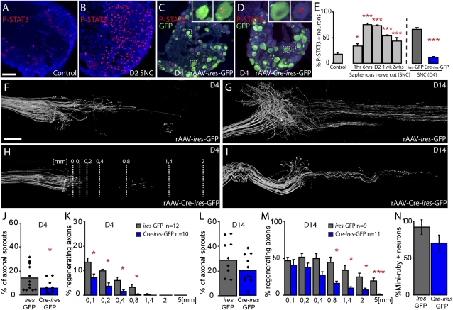
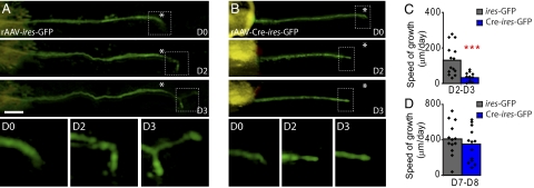
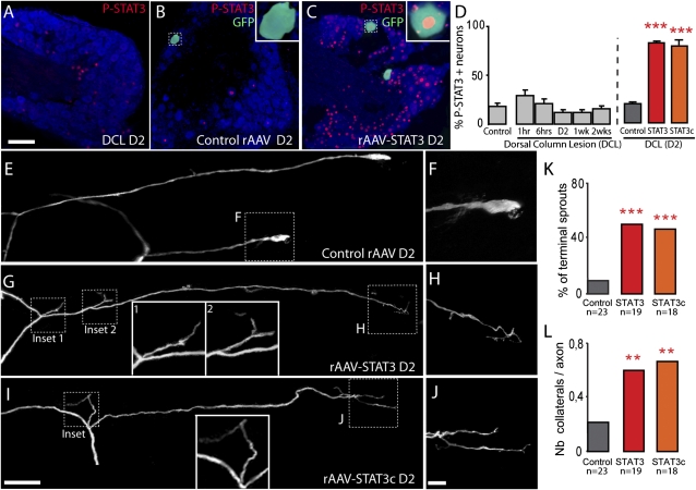
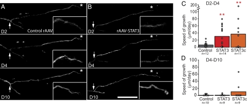
In the peripheral nervous system (PNS), damaged axons regenerate successfully, whereas axons in the CNS fail to regrow. In neurons of the dorsal root ganglia (DRG), which extend branches to both the PNS and CNS, only a PNS lesion but not a CNS lesion induces axonal growth. How this differential growth response is regulated in vivo is only incompletely understood. Here, we combine in vivo time-lapse fluorescence microscopy with genetic manipulations in mice to reveal how the transcription factor STAT3 regulates axonal regeneration. We show that selective deletion of STAT3 in DRG neurons of STAT3-floxed mice impairs regeneration of peripheral DRG branches after a nerve cut. Further, overexpression of STAT3 induced by viral gene transfer increases outgrowth and collateral sprouting of central DRG branches after a dorsal column lesion by more than 400%. Notably, repetitive in vivo imaging of individual fluorescently labeled PNS and CNS axons reveals that STAT3 selectively regulates initiation but not later perpetuation of axonal growth. With STAT3, we thus identify a phase-specific regulator of axonal outgrowth. Activating STAT3 might provide an opportunity to "jumpstart" regeneration, and thus prime axons in the injured spinal cord for application of complementary therapies that improve axonal elongation.


Conflict of interest statement

The authors declare no conflict of interest.

Figures

Fig. 1.
Fig. 1.
Deletion of STAT3 impairs regeneration of peripheral DRG axons after a saphenous nerve cut (SNC; A and B). Confocal images of L3 DRGs immunostained for P-STAT3 (red) and counterstained with fluorescent Nissl-like stain (NeuroTrace, cyan) in a control (unlesioned) WT mouse (A) and 2 d following an SNC (B). (C and D) L3 DRGs of STAT3fl/fl mice 4 d after SNC previously injected with rAAV-ires-GFP (C) or rAAV-Cre-ires-GFP (D; NeuroTrace, cyan; P-STAT3, red; GFP, green). GFP-positive DRG neurons (Insets, C and D) are at a magnification of ×3. (E) Quantification of the number of P-STAT3–positive DRG neurons (identified by NeuroTrace counterstaining) at different time points following SNC in WT mice and in STAT3fl/fl mice previously injected with rAAV-ires-GFP (gray column) or rAAV-Cre-ires-GFP (blue column) at 4 d following a SNC (n = 6 animals per group). (FI) Confocal images taken at 4 d (F and H) and 14 d (G and I) after SNC display the proximal stump of STAT3fl/fl saphenous nerves that receive fibers from L3 DRGs injected with control rAAV-ires-GFP (F and G) or rAAV-Cre-ires-GFP (H and I). (JM) Quantification of axonal sprouting at the site of lesion (J and L) and regeneration ratios (K and M) at different distances from the cut site (lines, H) of axons derived from STAT3-competent DRG neurons (ires-GFP, gray columns) and STAT3-deficient DRG neurons (Cre-ires-GFP, blue columns) at 4 d (J and K) and 14 d (L and M) after SNC. (N) Quantification of the percentage of L3 DRG neurons retrogradely labeled with the tracer Miniruby from distal STAT3-competent (ires-GFP, gray columns; n = 21 sections, n = 6 DRGs) and STAT3-deficient (Cre-ires-GFP, blue columns; n = 11 sections, n = 4 DRGs) saphenous nerves 28 d following SNC (values were normalized to the percentage of Miniruby-positive DRG neurons traced from the same anatomical localization in unlesioned mice). (Scale bars: A, 100 μm; F, 250 μm.)
Fig. 2.
Fig. 2.
In vivo imaging reveals that growth of STAT3-deficient axons is reduced in the initial but not the later stages of regeneration. (A and B) In vivo time-lapse images of saphenous nerve axons (GFP, green) in sparsely labeled animals emerging from DRGs that were injected with control rAAV-ires-GFP (A) or rAAV-Cre-ires-GFP (B). Axons were imaged immediately after transection (D0) as well as 2 and 3 d after transection (asterisk indicates lesion site, orange latex beads visible on the left were used as fiduciary markers). Lower row of boxes show the boxed axon tips at magnifications of ×3 over time. (C and D) Quantification of the growth speed of axons from STAT3-competent DRG neurons (ires-GFP, gray columns) and STAT3-deficient DRG neurons (Cre-ires-GFP, blue columns) during the early (C, 2–3 d after transection) and the late phase (D, 7–8 d after transection) of the regeneration process. (Scale bar: A, 100 μm.)
Fig. 3.
Fig. 3.
Viral vector gene transfer of STAT3 and STAT3c induces terminal and collateral sprouting of DRG branches after a central lesion. (AC) Confocal images of cervical DRGs immunostained for P-STAT3 (red) and counterstained with fluorescent Nissl-like stain (NeuroTrace, cyan) in a WT mouse 2 d following a dorsal column lesion (DCL, A) and in lesioned Thy1-GFPs mice (GFP, green) injected with control rAAV (B) or rAAV-STAT3 (C). Insets: Higher-magnification (×3) of the GFP-positive neurons boxed in the images. (D) Quantification of the number of P-STAT3–positive DRG neurons (identified by NeuroTrace counterstaining) at different time points following DCL in WT mice and in mice previously injected with control rAAV (gray column), rAAV-STAT3 (red column), or rAAV-STAT3c (orange column) at 2 d after a central lesion (n = 6 animals per group). (EJ) Confocal images of lesioned spinal axon endings derived from DRGs injected with control rAAV (bulbs, E and F), rAAV-STAT3 (terminal sprout, G and H), or rAAV-STAT3c (terminal sprout, I and J). (F, H, and J) Higher-magnification views of details boxed in E, G, and I. Additional insets (G and I) show a magnification ×2 of the boxed collateral sprouts. (K and L) Quantification of terminal (K) and collateral (L) sprouting of axons derived from DRGs injected with control rAAV, rAAV-STAT3, or rAAV-STAT3c and analyzed 2 d after transection. (Scale bars: A and I, 100 μm; J, 25 μm.)
Fig. 4.
Fig. 4.
In vivo imaging reveals successful initiation but not elongation of CNS axons after STAT3 and STAT3c gene therapy. (A and B) Multiphoton images of the growth pattern of spinal DRG axons emerging from DRGs injected with control rAAV (A) or rAAV-STAT3 (B) imaged 2, 4, and 10 d following a central lesion. Insets: Magnifications (×2) of boxed axon ends. (C and D) Quantification of axonal growth speed in vivo after injection of control rAAV (gray bars), rAAV-STAT3 (red bars), or rAAV-STAT3c (orange bars) analyzed early (C, 2–4 d) and late (D, 4–10 d) after transection. (Scale bar: B, 200 μm.)

References

    1. Schwab ME, Bartholdi D. Degeneration and regeneration of axons in the lesioned spinal cord. Physiol Rev. 1996;76:319–370. - PubMed
    1. Neumann S, Woolf CJ. Regeneration of dorsal column fibers into and beyond the lesion site following adult spinal cord injury. Neuron. 1999;23:83–91. - PubMed
    1. Ylera B, et al. Chronically CNS-injured adult sensory neurons gain regenerative competence upon a lesion of their peripheral axon. Curr Biol. 2009;19:930–936. - PubMed
    1. Raivich G, Makwana M. The making of successful axonal regeneration: Genes, molecules and signal transduction pathways. Brain Res Brain Res Rev. 2007;53:287–311. - PubMed
    1. Rossi F, Gianola S, Corvetti L. Regulation of intrinsic neuronal properties for axon growth and regeneration. Prog Neurobiol. 2007;81:1–28. - PubMed

Publication types

Substances