. 2011 Apr 1;71(7):2686-96.
doi: 10.1158/0008-5472.CAN-10-3513. Epub 2011 Mar 29.

Differentiation of NUT midline carcinoma by epigenomic reprogramming

Affiliations

Differentiation of NUT midline carcinoma by epigenomic reprogramming

Brian E Schwartz et al. Cancer Res. .

Abstract

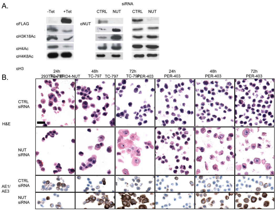
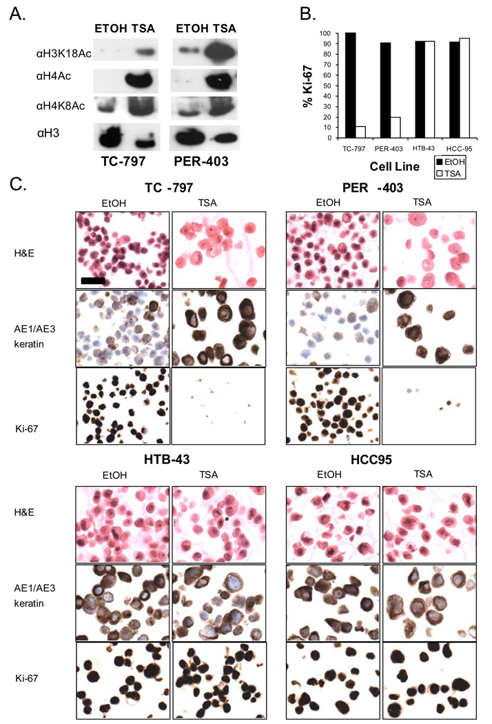
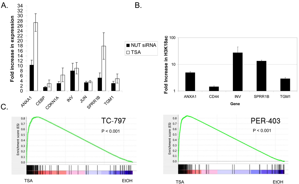
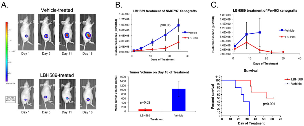
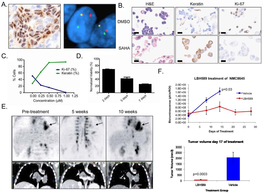
NUT midline carcinoma (NMC) is a lethal pediatric tumor defined by the presence of BRD-NUT fusion proteins that arrest differentiation. Here we explore the mechanisms underlying the ability of BRD4-NUT to prevent squamous differentiation. In both gain-of and loss-of-expression assays, we find that expression of BRD4-NUT is associated with globally decreased histone acetylation and transcriptional repression. Bulk chromatin acetylation can be restored by treatment of NMC cells with histone deacetylase inhibitors (HDACi), engaging a program of squamous differentiation and arrested growth in vitro that closely mimics the effects of siRNA-mediated attenuation of BRD4-NUT expression. The potential therapeutic utility of HDACi differentiation therapy was established in three different NMC xenograft models, where it produced significant growth inhibition and a survival benefit. Based on these results and translational studies performed with patient-derived primary tumor cells, a child with NMC was treated with the FDA-approved HDAC inhibitor, vorinostat. An objective response was obtained after five weeks of therapy, as determined by positron emission tomography. These findings provide preclinical support for trials of HDACi in patients with NMC.


Figures

Figure 1
Figure 1
BRD4-NUT expression is associated with blocked differentiation, and repression of histone acetylation. (A) Effects on histone acetylation resulting from induced expression of flag-BRD4-NUT in 293TRex cells, and siRNA-induced knockdown of BRD4-NUT in two NMC cell lines after 24 hr.. (B) siRNA-induced knockdown of BRD4-NUT using an siRNA targeting NUT in two NMC cell lines. Hematoxylin and eosin staining (H & E) as well as the immunohistochemical (IHC) detection of keratin with antibody AE1/AE3 are shown for each cell line 24h, 48h, and 72h following knockdown. Magnification (scale bar, 25um) is identical for all panels.
Figure 2
Figure 2
Treatment of NMC cells with the HDACi, trichostatin A, restores global histone acetylation and induces squamous differentiation and arrested growth. (A) Changes in histone acetylation resulting from TSA treatment of two NMC cell lines after 24 hr. (B) Ki-67 (marker of cycling cells) fraction in two NMC cell lines (TC-797 and PER-403) and two non-NMC squamous cell carcinoma cell lines (HTB-43 and HCC-95). Value is from a denominator of 200 viable cells counted. (C) Morphologic and immunophenotypic changes following TSA (72h, 25nM) treatment of NMC cell lines, TC-797, PER-403, and non-NMC cell lines, HTB-43 and HCC-95. Magnification (scale bar, 25um) is identical for all panels.
Figure 3
Figure 3
Histone deacetylase inhibition induces a very similar program of differentiation with that following knockdown of BRD4-NUT in NMC cells. (A) Quantitative RT-PCR analysis of seven selected squamous differentiation-specific genes 24h following siRNA-knockdown and TSA (25nM) treatment of TC-797 NMC cells. Results are from triplicate independent samples. ct, control scrambled siRNA. (B) Quantitative PCR of ChIPd DNA from NMC TC-797 cells using primers to the promoter regions of select differentiation specific genes (from A, above), using H3K18Ac antibody. Values, obtained from independent duplicate samples, are relative to IgG ChIP control. (C) Gene set enrichment analysis (GSEA) measuring the correlation of genes up-regulated following knockdown of BRD4-NUT in both TC-797 and PER-403 NMC cell with HDACi-mediated expression changes. In these plots, vertical lines indicate the rank order of the knockdown gene set genes within the HDACi-treated cells (top: TC-797, bottom: PER-403). Red (left): genes up-regulated by TSA treatment; blue (right) down-regulated genes. The concentration of vertical lines within the TSA (red) portion of the spectrum reflected in the running enrichment score plot (green line) indicates the degree of correlation between up-regulation in response to BRD4-NUT knockdown or TSA treatment.
Figure 4
Figure 4
Growth inhibition of xenograft models of two NMCs by the HDACi LBH-589. (A) Representative bioluminescence images of TC-797 xenografts treated with vehicle or LBH589 at 10 mg/kg IP. (B) Effects of LBH589 treatment on TC-797 (n=6 per group) xenograft growth measured by bioluminescence and tumor volume. (C) Bioluminescence imaging and survival analysis of PER-403 xenografts (n=5 per group) treated with LBH589. Mice were sacrificed when tumors reached 2 cm. Due to rapid tumor growth, the bioluminescence data for vehicle-treated animals (upper panel) is confounded by the need to sacrifice animals bearing large tumors beginning on treatment day 18 (lower panel).
Figure 5
Figure 5
Morphologic and immunophenotypic squamous differentiation correlates with increased acetylation of TC-797 xenografts treated with HDACi (LBH-589). AcL, immunohistochemistry using anti-acetyl-lysine antibody (Cell Signaling Technologies); Veh1, vehicle control (DMSO) treated mouse 1; Veh2, vehicle treated mouse 2; LBH1, LBH-589 treated test mouse 1; LBH2, LBH-589 treated test mouse 2. Magnification (scale bar, 25um) is identical for all panels.
Figure 6
Figure 6
Diagnosis and response of an NMC patient to single-agent treatment with the HDAC inhibitor vorinostat (SAHA). (A) Diagnosis of NMC in a 10 year old boy by immunohistochemical (IHC) staining with a monoclonal antibody to NUT (27) and by dual color, split-apart fluorescence in situ hybridization (FISH) (35) using probes flanking the NUT locus. (B) Morphologic and immunophenotypic effects of 72h treatment with SAHA (1µM) versus DMSO control. IHC was performed for keratins (AE1/AE3 antibodies) and the proliferation marker, Ki-67. (C) Dose-response curve of the patient’s tumor cells to SAHA (IC50=250 nM), as measured by Ki-67 fraction and keratin-positivity. (D) Cell growth in SAHA (1 µM), as measured by ATP content relative to DMSO control. (E) P18PF-fluorodeoxyglucose–positron emission tomography (FDG-PET) and CT scan of the patient’s mediastinal tumor (arrow). (F) Growth response of NMC 8645 xenografts (n=6 vehicle, and n=7 LBH589 groups) derived from this patient as measured by bioluminescence and tumor volume.

References

    1. Druker BJ, Tamura S, Buchdunger E, et al. Effects of a selective inhibitor of the Abl tyrosine kinase on the growth of Bcr-Abl positive cells. Nature medicine. 1996;2(5):561–566. - PubMed
    1. Cools J, DeAngelo DJ, Gotlib J, et al. A tyrosine kinase created by fusion of the PDGFRA and FIP1L1 genes as a therapeutic target of imatinib in idiopathic hypereosinophilic syndrome. The New England journal of medicine. 2003;348(13):1201–1214. - PubMed
    1. Di Croce L, Raker VA, Corsaro M, et al. Methyltransferase recruitment and DNA hypermethylation of target promoters by an oncogenic transcription factor. Science (New York, NY. 2002;295(5557):1079–1082. - PubMed
    1. Redner RL, Corey SJ, Rush EA. Differentiation of t(5;17) variant acute promyelocytic leukemic blasts by all-trans retinoic acid. Leukemia. 1997;11(7):1014–1016. - PubMed
    1. Soignet SL, Maslak P, Wang ZG, et al. Complete remission after treatment of acute promyelocytic leukemia with arsenic trioxide. The New England journal of medicine. 1998;339(19):1341–1348. - PubMed

Publication types

MeSH terms